
J. Morita
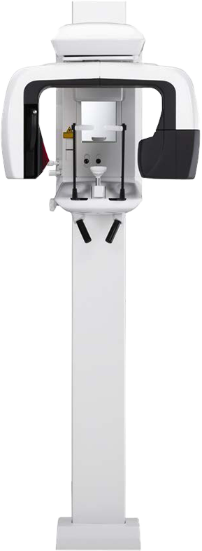
Veraviewepocs (X550) 3D R100
Multifunctional J. Morita unit delivering high resolution, 125 µm voxel images with panoramic and cephalometric imaging.
Serving dentistry since 2003
Installation, training & expert support included
About the Veraviewepocs (X550) 3D R100
High resolution, 125 µm voxel images provide a clear observation of the periodontal pocket, the periodontal membrane, and the alveolar bone, as well as soft tissue such as the maxillary sinus membrane, with a 3D volume captured in about 9.4 seconds and resolution better than 2 line pairs per mm (MTF 10%). A special dose reduction mode lowers exposure for soft tissue which reduces dosage to a mere 60% of the standard mode. The Veraviewepocs 3D R100 is a distinct J. Morita product line from the Veraview X800 platform, built around two features unique to this unit. Panoramic Scout lets you mark the region of interest directly on a panoramic image on screen, and the C-arm repositions itself automatically to center the 3D capture there. The 3D Reuleaux Full Arch FOV replaces the usual cylindrical volume with a convex-triangle shape that follows the natural curve of the arch, covering the full maxilla or mandible at a lower dose than a same-diameter cylindrical scan would require. The R100 won J. Morita's Innovations Award in 2012. Multifunctional, it also offers panoramic and cephalometric imaging in the same unit — a lateral cephalometric capture runs 2.6 to 5.8 seconds with roughly a tenth the dose of a conventional film-based X-ray, and image processing finishes in about 20 seconds. With a total of 6 fields of view, 3D R100 is ideal for implantology as well as being suitable for periodontology, orthodontics, endodontics, oral surgery, and general dentistry.
- FOVs: Ø 40 x H 40 mm, Ø 40 x H 80 mm, Ø 80 x H 50 mm, Ø 80 x H 80 mm; 3D Reuleaux Full Arch FOVs: Ø 100 x H 50 mm, Ø 100 x H 80 mm
- Minimum voxel size: 125 µm
- Dose reduction mode lowers soft-tissue exposure to 60% of the standard mode
- Panoramic and cephalometric imaging in one unit
- Focal spot: 0.5mm
- Software: i-Dixel, Exoplan (optional)
- Space requirements: 40.15" x 51.18" Pan only, or 78.74" x 51.18" with Cephalometric (Width x Depth)
- 3 Year Manufacturer Warranty on Parts
- 3D exposure time: approximately 9.4 seconds
- Resolution greater than 2 line pairs/mm (MTF 10%)
- Panoramic Scout: mark the region of interest on a panoramic image and the C-arm repositions automatically for the 3D capture
- Two-direction scout imaging plus 5-beam laser positioning, with automatic C-arm repositioning to confirm alignment
- Partial panoramic function excludes up to 5 sections plus the maxillary sinus area to reduce dose
- Cephalometric lateral projection: 2.6-5.8 seconds capture time, image processing in about 20 seconds, with 3 selectable partial cephalometric patterns to reduce dose
- Won J. Morita's Innovations Award in 2012
New to CBCT, or narrowing down field of view, voxel size, and cost? Read the complete guide to dental CBCT before you decide.
Under the hood
Veraviewepocs (X550) 3D R100 technology
Panoramic Scout
Before the 3D exposure, the unit captures a high-resolution panoramic image and displays it on the PC monitor so the region of interest can be marked directly on that image rather than estimated from external reference points. The C-arm then repositions itself automatically to center the 3D capture on that spot, and a two-directional scout confirms the position before the CBCT volume is taken. It's a faster, more repeatable way to line up an implant site or an impacted tooth than manual laser alignment alone.
3D Reuleaux Full Arch FOV
Instead of a cylindrical volume, the Reuleaux Full Arch field of view is shaped like a convex triangle that follows the natural curve of the dental arch, available at Ø100mm (equivalent) in 50mm or 80mm heights. By excluding the corner volume a cylinder would otherwise capture outside the arch, it covers the full maxilla or mandible at a lower dose than a same-diameter cylindrical scan would require. That makes full-arch implant planning and oral surgery cases practical without stepping up to a larger, higher-dose FOV.
Autofocus (AF Automatic Positioning)
A light-beam sensor measures the distance to the patient's teeth and automatically moves the arm into the optimal position for a panoramic exposure, without the patient needing to be repositioned by hand. Because the same measurement runs every time, image layer positioning is more reproducible chair to chair and operator to operator, which cuts down on retakes from an off-center focal trough.
Digital Direct Auto Exposure (DDAE)
DDAE monitors the sensor's signal in real time during the panoramic exposure and adjusts X-ray output on the fly to match the density of the anatomy the beam is passing through at that instant, rather than relying on a single preset exposure for the whole arch. That produces a wider dynamic range across the image, so dense areas like the symphysis and thin areas like the ramus both come out readable in the same exposure.
Auto Image Enhancement-High Definition (AIE-HD)
AIE-HD is a software processing step that applies a logarithmic conversion to the captured panoramic image, evening out overall density and pulling out detail in the shaded and overexposed regions a raw exposure tends to lose. It runs automatically after capture, so the image on screen is already balanced for reading rather than needing manual contrast adjustment first.
Dose Reduction Mode
Dose Reduction Mode lowers tube output for exposures where the anatomy doesn't need full-strength penetration, cutting dose to about 60% of the standard mode setting on Ø40 x H80mm captures. Morita's own comparison shows soft-tissue structures like the maxillary sinus membrane rendering with fewer artifacts in this mode than in a standard exposure, which makes it a reasonable choice when the exam is focused on soft tissue rather than dense bone.
Veraviewepocs (X550) 3D R100 questions, answered
What field of view does the J. Morita Veraviewepocs 3D R100 have?
The J. Morita Veraviewepocs 3D R100 offers fields of view from Ø 40 x H 40 mm up to Ø 100 x H 80 mm, including 3D Reuleaux Full Arch options at Ø 100 mm, and its minimum voxel size is 125 µm.
Is the Veraviewepocs 3D R100 good for implants and endodontics?
The Veraviewepocs 3D R100 is ideal for implantology and is also suitable for periodontology, orthodontics, endodontics, oral surgery, and general dentistry. Its 125 µm voxel images provide a clear view of the periodontal pocket, periodontal membrane, alveolar bone, and soft tissue such as the maxillary sinus membrane.
Does the Veraviewepocs 3D R100 include panoramic and cephalometric imaging?
Yes, the Veraviewepocs 3D R100 is a multifunctional unit that combines CBCT with panoramic and cephalometric imaging in one machine.
How does the Veraviewepocs 3D R100 reduce patient dose?
The Veraviewepocs 3D R100 has a special dose reduction mode that lowers exposure for soft tissue, reducing dosage to 60% of the standard mode.
What software does the Veraviewepocs 3D R100 work with?
The Veraviewepocs 3D R100 works with i-Dixel and optional Exoplan software. Every scan also exports in the standard DICOM format, so it's compatible with any DICOM-compliant viewing or planning software your practice already uses, and Dental TI integrates it as part of installation.
What is included when I buy the Veraviewepocs 3D R100 from Dental TI?
Every Veraviewepocs 3D R100 purchase from Dental TI includes site planning and installation, integration with your practice's imaging software, on-site team training (remote also available), and expert remote and on-site support. Financing is available, and the unit carries a 3 year manufacturer warranty on parts.
What is Panoramic Scout and how does it simplify positioning?
Panoramic Scout captures a panoramic image first and lets you mark the region of interest directly on that image on screen, instead of estimating the position from external landmarks. The C-arm then repositions itself automatically to center the 3D capture on the marked spot, and a two-directional scout confirms the alignment before the CBCT exposure is taken.
What does the Reuleaux full-arch field of view do differently from a standard cylindrical CBCT volume?
Instead of a cylinder, the 3D Reuleaux Full Arch FOV is shaped like a convex triangle that follows the natural curve of the dental arch. That shape covers the full maxilla or mandible (Ø100mm equivalent, in 50mm or 80mm heights) while excluding the extra corner volume a same-diameter cylindrical scan would otherwise capture, which is how it holds dose down on a full-arch exposure.
How much radiation dose does the cephalometric scan use compared to conventional X-ray?
A lateral cephalometric capture on the Veraviewepocs 3D R100 uses roughly a tenth of the dose of a conventional film-based X-ray, per Morita's own comparison, with a capture time of 2.6 to 5.8 seconds and image processing finishing in about 20 seconds. A partial cephalometric option with 3 selectable patterns can reduce dose further when a full lateral image isn't needed.
How fast is a 3D scan on the Veraviewepocs 3D R100, and how sharp is the image?
A standard 3D exposure takes about 9.4 seconds, and resolution runs greater than 2 line pairs per mm at MTF 10% across the unit's 125 µm minimum voxel size. That combination is what gives the R100 clear detail on the periodontal pocket, periodontal ligament, and alveolar bone that implant planning and periodontal cases depend on.
Has the Veraviewepocs 3D R100 won any industry recognition?
Yes. The Veraviewepocs 3D R100 won J. Morita's Innovations Award in 2012, the year it launched, largely on the strength of its patent-pending 3D Reuleaux Full Arch FOV.
Is the Veraviewepocs (X550) 3D R100 right for your practice?
Talk it through with an advisor who installs these machines every week — including the ones we'd steer you away from.