
CBCT 201: Understanding Slice Thickness
· Dental TI
Understanding how to vary slice thickness and why and when you want to do that. I realized that for those that are new to CBCT the concept of slice thickness is foreign and needs further explanation.
Voxel Size and the Thinnest Slice
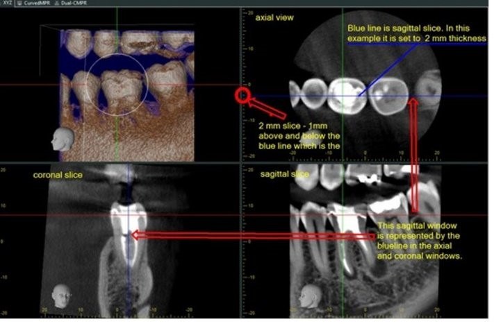
First off, the thinnest slice is the voxel size that was used to acquire the image. A voxel is a cube and a CBCT volume is compiled of thousands of voxels. The slice appears in the axial, coronal and sagittal views. The following illustration explains this concept. The sagittal (lower right window side view) corresponds to the blue line that is in the axial view and coronal view. With a 2mm slice thickness you have a composite image that is comprised of slices. 1mm above and below the blue line in the axial window. The thinnest slice possible is 1 voxel which in this case is .125mm. This means we have combined 16 .125mm slices to have a thickness of 2mm. (16X.125 = 2).
Comparing Slice Thickness in the Sagittal View
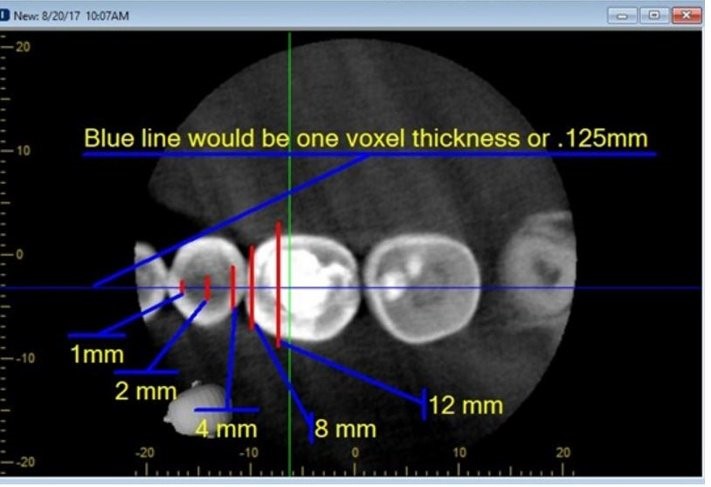
The following axial image illustrates the difference in thickness of the sagittal images that follow:
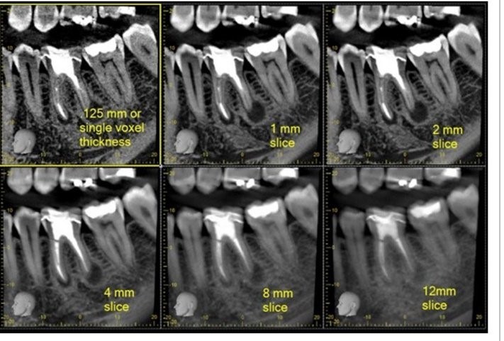
Sagittal images corresponding to thicknesses presented in the above image:
When to Vary Slice Thickness
By increasing slice thickness you will see a composite view that becomes very similar (although it does not have the same line pair resolution) to a P.A. Often time varying the thickness gives the observer a composite look that is easier to understand than a single slice or even 1mm view. Understanding slice thickness and when to vary it gives the CBCT interpreter a different look at the information that can lead to a more certain diagnoses. At the largest thickness the CBCT reader is presented with an image that looks much like a traditional PA while dropping the slice thickness allows for the observation of smaller details and their exact location.