
A Different Perspective: MPR-Curves in i-Dixel
· Dental TI
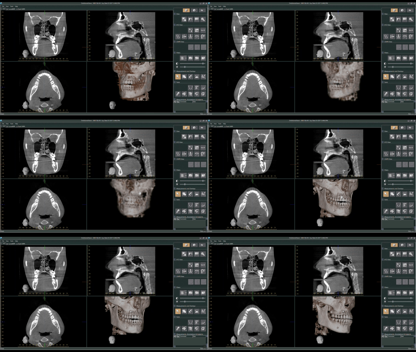
J. Morita’s 3D viewing and treatment planning software, i-Dixel, offers a robust set of tools to review scans and plan treatment digitally. When a scan is loaded, the data is presented in the “volume view” with windows for axial, coronal, sagittal, and 3D perspectives:
The MPR (Multi-Planar Reconstruction) and Dual-MPR tabs allow for different (and arguably more precise) perspective views to analyze a CBCT volume and plan treatment. In either MPR tab, the user draws an “MPR line/curve”, along which the software places perpendicular “slices” at adjustable intervals which allow the user to progress through cross-sections of the patient’s anatomy in a slice-by-slice perspective. This is a useful tool— for example— to analyze discrete regions of interest, trace the mandibular canal for implants, and verify the angle and depth of a planned implant.
Below, we will explore some of the different views that can be created by tracing an MPR curve.
Extracting a 2D Pan from a 3D Volume
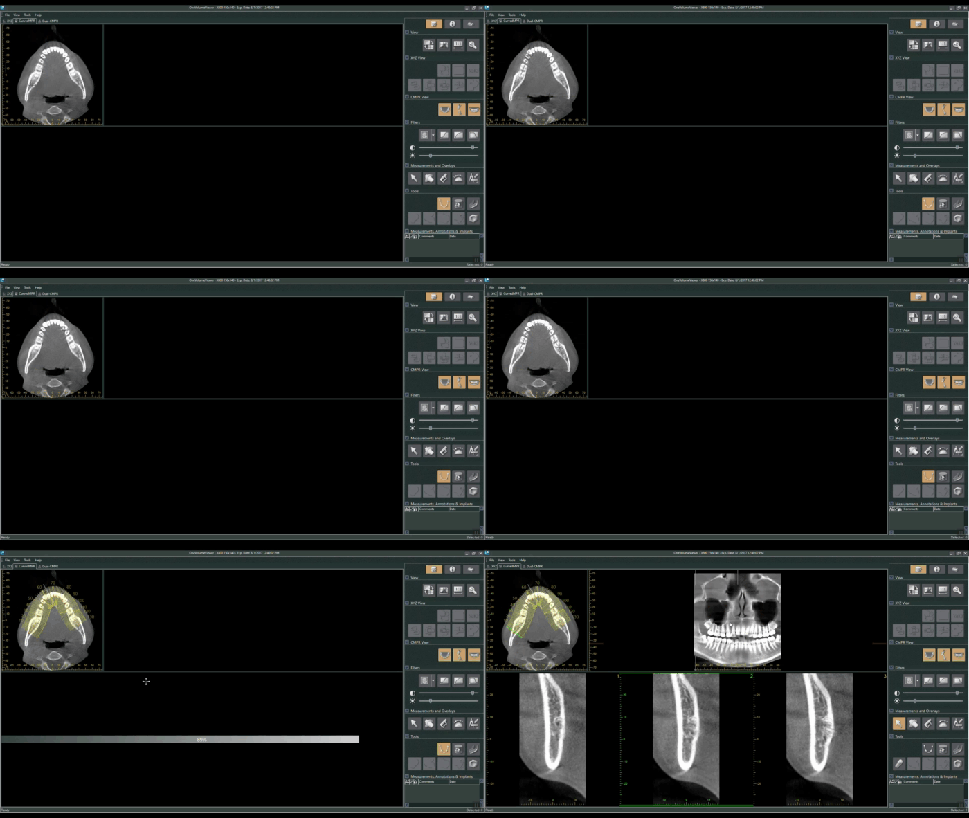
By tracing our MPR curve along the entire arch, the user is able to create an extracted panoramic image from their 3D volume. To the right of the axial view (in which the MPR curve is drawn), the software compiles a flattened or panoramic image of whatever anatomy the MPR curve spans. If one draws a curve that spans the entire arch, the software will give a panoramic view of the patient’s mouth.
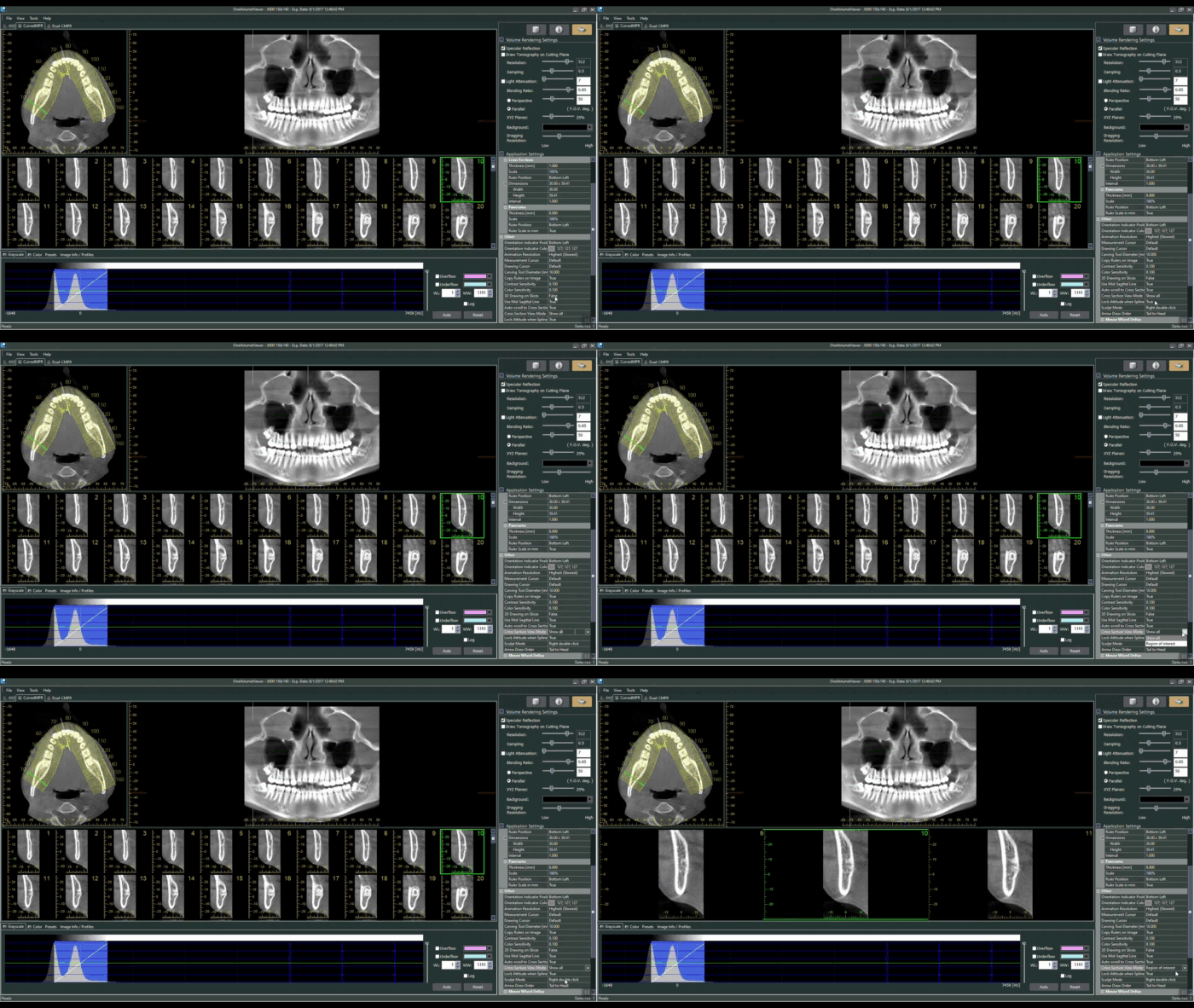
*Useful Tip* The number of slices displayed in the bottom window can be adjusted by changing the “Cross Section View Mode” under the Settings section of the viewer:
Mesial-Distal
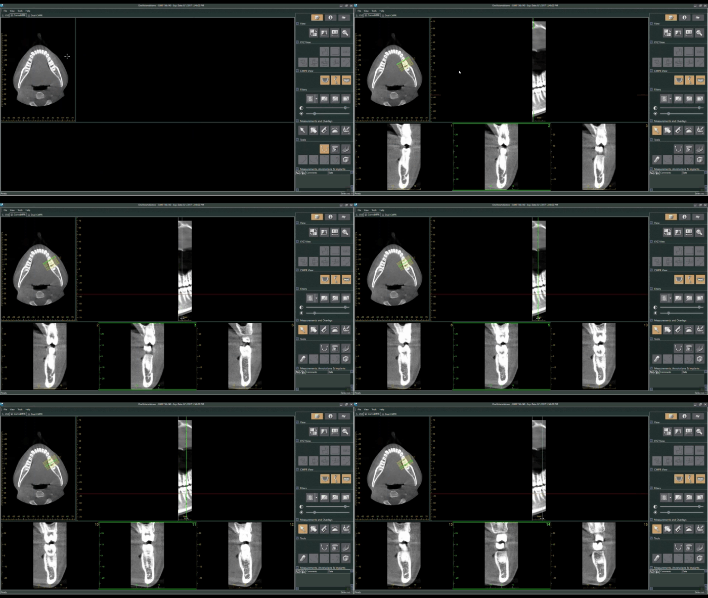
Tracing an MPR-Curve along a single tooth, implant site, or a few teeth, will provide the user with coronal-perspective set of slices to progress through the mesial-distal aspects of the anatomy. This view is especially useful for mapping the mandibular canal for lower-arch implant cases.
Buccal-Lingual
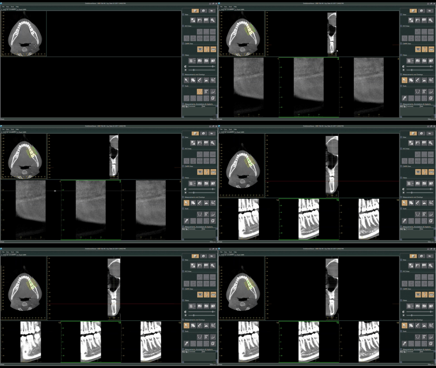
Tracing an MPR line across a tooth or implant site from the buccal to lingual side will allow the user to progress through the area from a sagittal perspective:
Radial
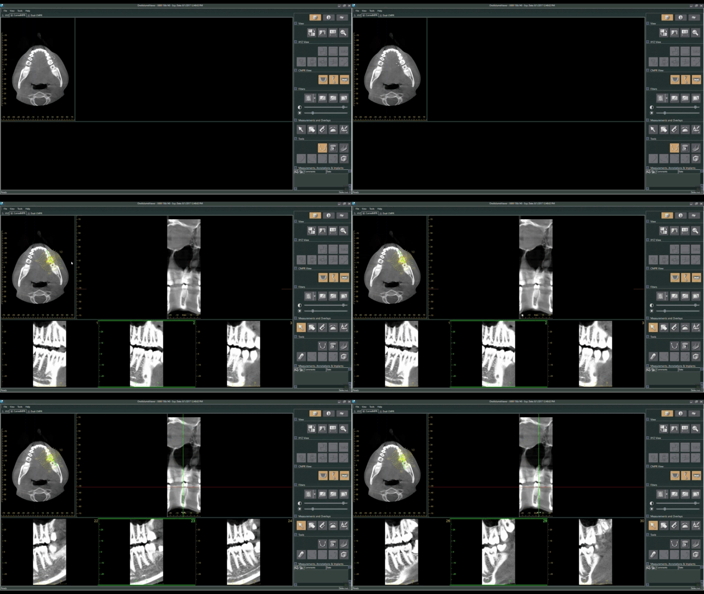
The MPR curve can also be drawn into a shape. Creating a circle around a tooth will allow the user to examine circumferential aspects of the targeted area:
Root-Crown (Long Axis)
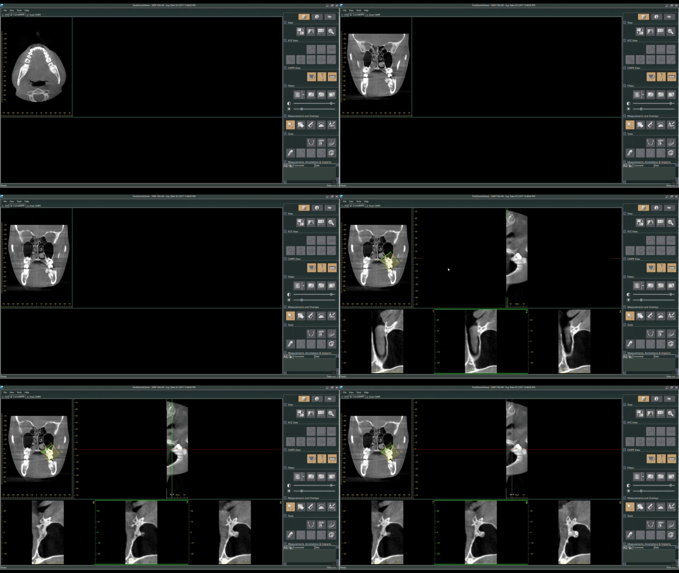
The user is also able to rotate the axial view in the top-left window to a coronal perspective by clicking and dragging their mouse cursor. Drawing the MPR line from a tooth’s root to its crown in this view will give us axial slices to progress through. This view can be very useful for locating missed canals, or examining teeth/implants that may have issues related to the patient’s sinus cavity.
Dual-MPR
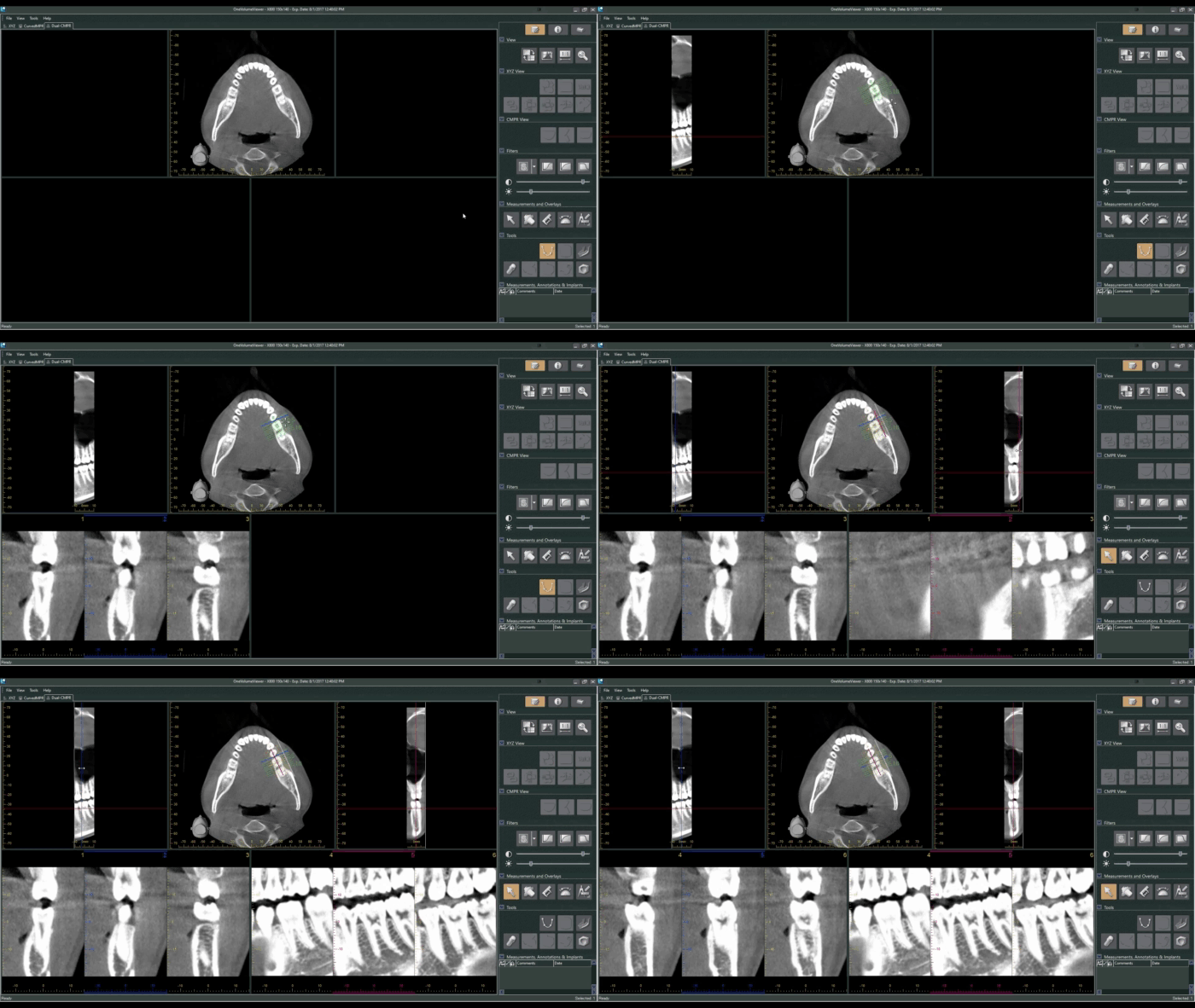
The Dual-MPR tab allows the user to draw two MPR lines for two different slice-by-slice perspectives. Below, we draw a mesial-distal and buccal-lingual MPR curve to allow us to view the tooth through both coronal and sagittal slices. The Dual-MPR function is especially ideal for verifying the depth and angle of a planned implant from multiple perspectives.
MPR curves can provide a more focused view of anatomical landmarks, and allow the user to create more diagnostic images for case presentation. For planning implants and mapping the mandibular canal, the MPR or Dual-MPR view is usually preferable to the volume view as they allow for a more precise view of the specific area.
Dental TI clients can connect with our staff for free one-on-one or team training on volume review and treatment planning functions in i-Dixel. Not a client? Feel free to request a free consultation to learn more about how Dental TI can help you get the most out of your imaging technology.