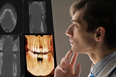
Understanding the Benefits of the CBCT Machine in Dental Care
· David Hanning
 CBCT is a Valuable Tool for Dentistry
Key Takeaways
- CBCT machines use cone-shaped X-ray beams to capture 150-400 images in 10-40 seconds, creating detailed 3D models of dental structures
- Modern CBCT scanners deliver 200-300 times less radiation than traditional CT while providing superior diagnostic accuracy.
- Essential applications include dental implant planning, orthodontics, endodontics, and complex oral surgery procedures.
- Investment in CBCT technology typically pays for itself through expanded service offerings such as dental implants and improved treatment outcomes.
- Leading manufacturers like J.Morita, Planmeca, HDXWill, Carestream and others offer models ranging from compact units to full-featured systems
The landscape of dental imaging has undergone a revolutionary transformation with the introduction of cone beam computed tomography technology. Modern dental practices are increasingly recognizing that CBCT machines represent far more than just another piece of equipment; they’re strategic investments that fundamentally change how clinicians diagnose, plan, and execute treatments across virtually every dental specialty.
For dental professionals considering this technology, understanding the capabilities, applications, and investment implications of a CBCT machine becomes crucial for making informed decisions that will shape their practice for years to come.
This comprehensive guide examines everything from the technical principles underlying cone beam CT to the practical considerations of selecting and implementing the right system for your specific needs.
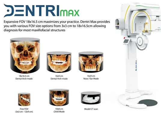 HDXWill DentriMax CBCT and FOVs
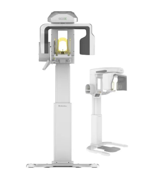
What is a CBCT Machine?
A CBCT machine, or cone beam computed tomography scanner, represents a specialized form of medical imaging technology specifically designed to produce three-dimensional volumetric images of dental and craniofacial structures. Unlike traditional dental x ray equipment that captures flat, two-dimensional images, cbct imaging creates detailed 3D models that reveal anatomical relationships previously invisible to clinicians.
The technology employs a cone-shaped X-ray beam that rotates 360 degrees around the patient’s head, capturing between 150 and 200 high-resolution 2D projection images during a single scan cycle. This cone beam CT exam typically requires only 10-40 seconds of scanning time, depending on the selected field of view and specific imaging protocol.
What distinguishes dental cone beam CT from conventional medical CT scanners is its optimization for oral and maxillofacial applications. While traditional CT scanners were designed for whole-body imaging with emphasis on soft tissue contrast, CBCT technology prioritizes the visualization of dental structures, bone density assessment, and precise anatomical measurements required for dental treatment planning.
The CBCT scanner generates what’s known as a volumetric data set—a digital representation of the scanned anatomy that can be manipulated, measured, and viewed from any angle. This capability transforms how dental professionals approach complex cases, offering unprecedented insight into tooth orientation, nerve paths, bone structure, and spatial relationships between anatomical landmarks.
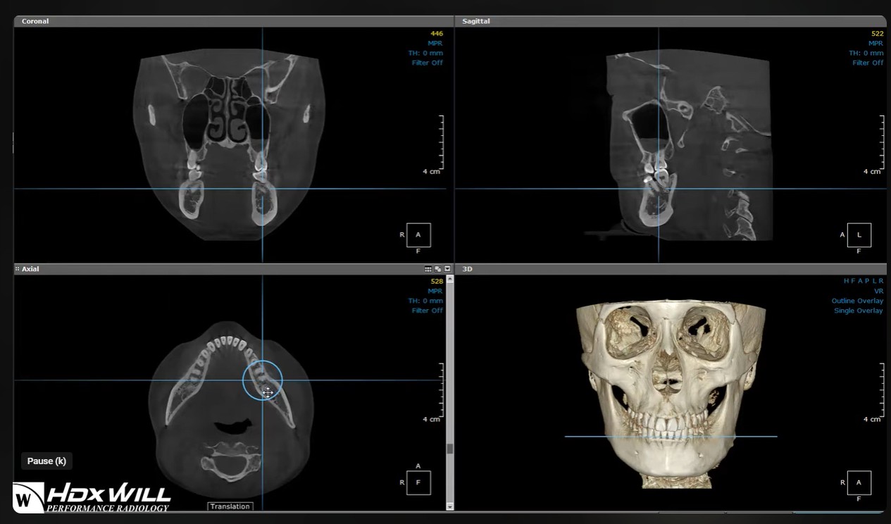 HDX Will Master Software Illustrates 3D Volume
How CBCT Machines Work
The fundamental principle behind CBCT technology involves the coordinated movement of an X-ray source and detector positioned on opposite sides of a revolving C-arm or gantry system. As this assembly rotates around the patient, the cone beam ct scanner captures hundreds of projection images from different angles, creating the raw data necessary for three-dimensional reconstruction.
The cone-shaped X-ray beam differs significantly from the fan-shaped beam used in conventional CT scanners. This configuration enables the CBCT machine to capture a substantially larger volume of data in a single rotation, thereby reducing scanning time while maintaining high spatial resolution. The beam geometry also contributes to the reduced radiation exposure, making cone beam CT imaging safer for routine dental applications.
Advanced reconstruction algorithms process these multiple 2D projections into detailed 3D volumetric data through a process called back-projection. Modern CBCT systems can achieve voxel resolution as acceptable as 0.076mm, providing exceptional detail for determining bone structure, nerve canals, and other critical anatomical features essential for precise treatment planning.
Field of view options represent another crucial aspect of how CBCT machines operate. Practitioners can select from small regional scan areas as small as 30x30 mm for focused endodontic applications, up to full-head scans encompassing 180x165 mm for comprehensive oral and maxillofacial evaluation.
This flexibility enables clinicians to minimize radiation exposure while capturing the exact anatomical information required for each specific case.
The digital workflow integration ensures that CBCT images are immediately available for diagnosis and treatment planning. Unlike traditional X-ray procedures that required film processing, cone beam CT imaging provides instant access to diagnostic data, streamlining patient care and reducing appointment times.
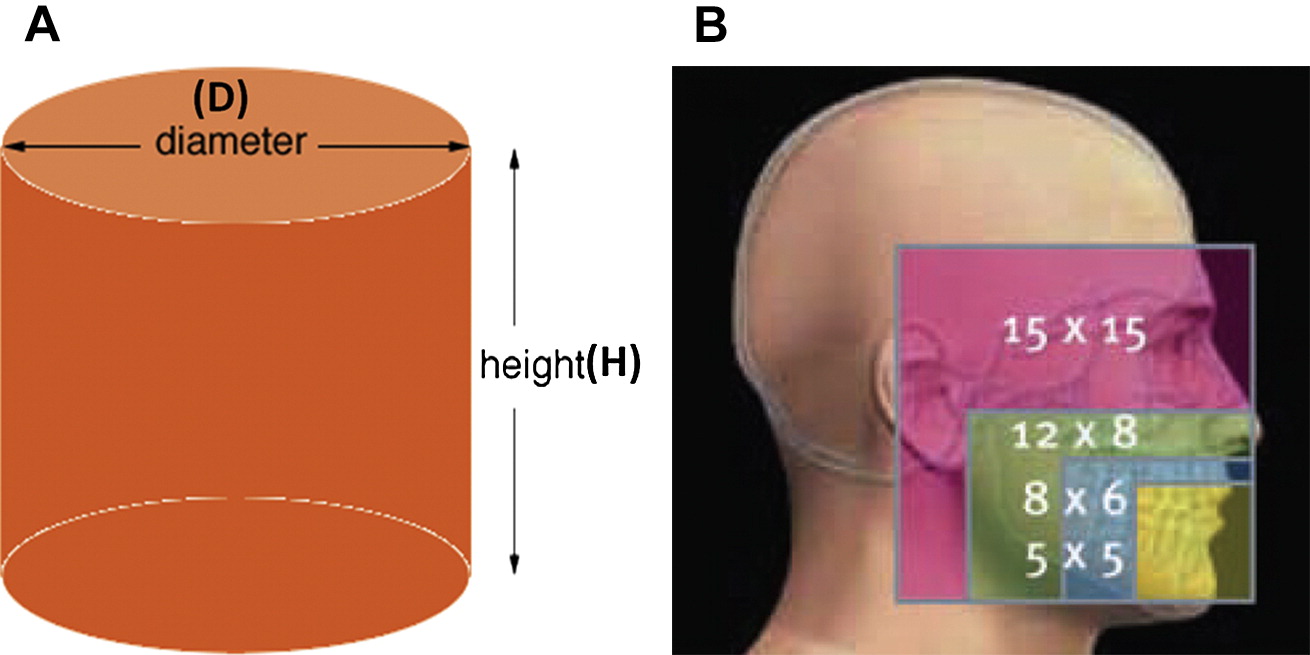 Illustration of Different FOVs
Essential CBCT Applications in Modern Dentistry
Dental Implant Planning
Dental implant success depends heavily on precise placement planning, making CBCT imaging an indispensable tool for implant dentistry. The technology enables comprehensive bone density assessment and volume measurements that determine implant site suitability and optimal positioning strategies.
During dental implant planning, CBCT scans provide critical information about available bone height and width, proximity to vital structures such as the mandibular nerve and maxillary sinus, and bone quality characteristics that influence implant stability. This three-dimensional perspective allows for virtual implant placement before surgery, significantly reducing complications and improving long-term success rates.
Modern CBCT systems integrate seamlessly with CAD/CAM technology and surgical guide fabrication, enabling computer-guided implant placement with sub-millimeter accuracy. This integration supports more precise treatment planning and execution, particularly valuable for full-arch restorations and complex reconstructive procedures.
The American Dental Association recognizes CBCT imaging as the standard of care for dental implant evaluation, citing its superior diagnostic capabilities compared to traditional dental X-ray methods. Studies consistently demonstrate improved implant success rates and reduced surgical complications when cone beam CT is utilized for comprehensive pre-surgical assessment.
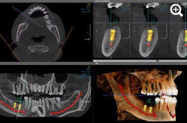 CBCT Used for Precise Implant Planning
Orthodontic Treatment Planning
Orthodontic applications of CBCT technology extend far beyond simple tooth positioning analysis. Modern orthodontists utilize cone-beam CT imaging for comprehensive 3D cephalometric analysis, airway assessment, and evaluation of craniofacial growth patterns that inform treatment approaches.
Root position evaluation becomes particularly critical before initiating tooth movement, especially in cases involving impacted teeth or previously traumatized areas. CBCT imaging reveals root morphology and proximity relationships that help orthodontists plan safe, predictable tooth movement while avoiding root damage or periodontal complications.
Temporomandibular joint analysis represents another significant application area. The detailed visualization of joint structures, condylar positioning, and articular disc relationships supports accurate diagnosis of TMJ disorders and guides appropriate treatment interventions.
Treatment progress monitoring through follow-up CBCT scans allows orthodontists to assess bone remodeling, root positioning changes, and overall treatment response. This capability proves especially valuable in complex cases involving surgical orthodontics or significant skeletal discrepancies.
Watch: 3D cephalometric tracing in HDXWill’s orthodontic analysis software
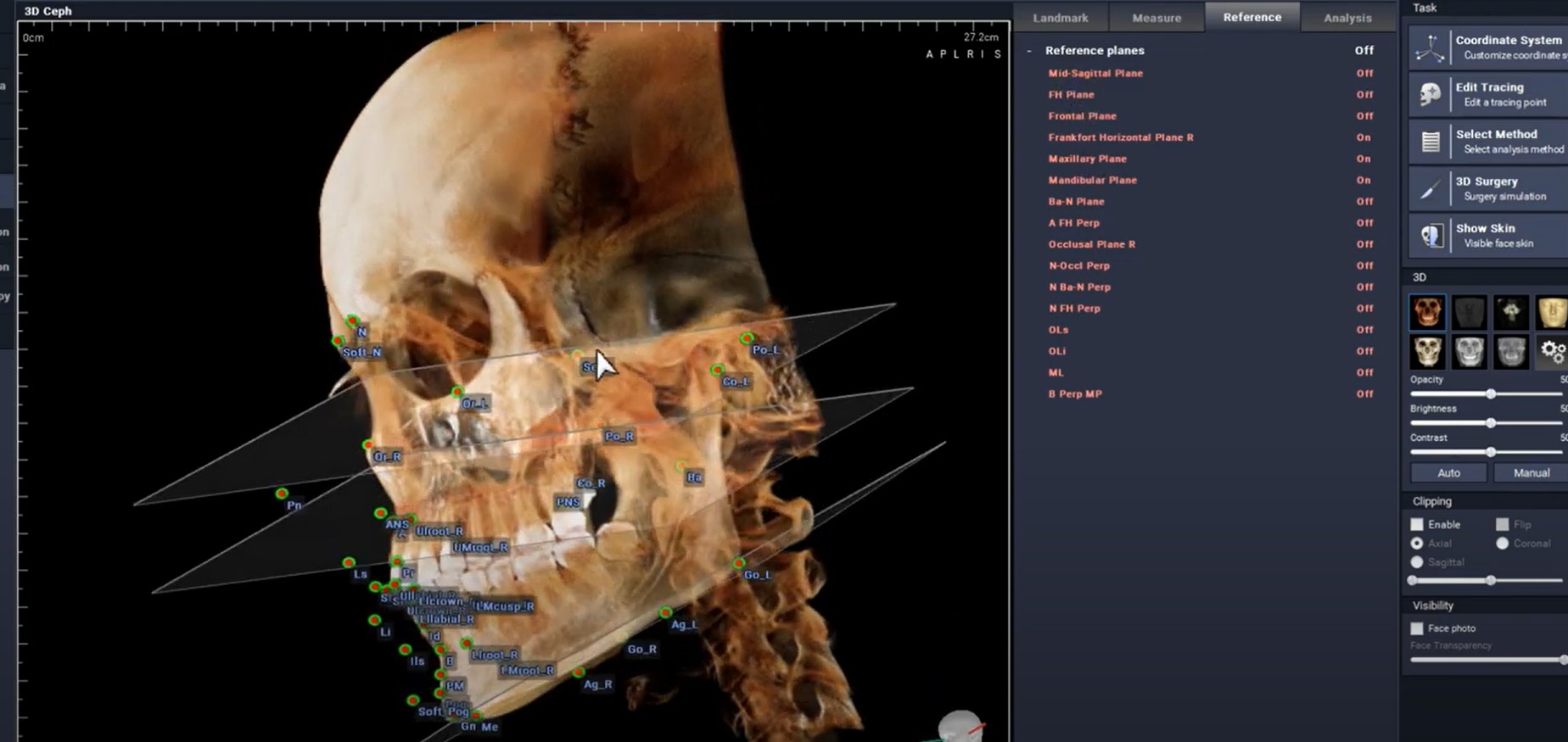 3D Ceph Tracing in HDXWill’s Orthodontic Analysis Software
Endodontic Procedures
Endodontic treatment success often depends on the clinician’s ability to locate and thoroughly clean all root canal systems. CBCT imaging revolutionizes endodontic diagnosis and treatment planning by revealing root canal morphology details invisible on traditional dental X-ray images.
Complex root canal anatomy, including additional canals, canal connections, and anatomical variations, becomes clearly visible through cone beam ct imaging. This enhanced visualization is particularly valuable in posterior teeth, where complex anatomy frequently contributes to endodontic treatment failures.
Periapical lesion assessment and treatment monitoring benefit significantly from the three-dimensional perspective provided by CBCT scans. The technology enables accurate measurement of lesion size, assessment of cortical plate involvement, and monitoring of healing progress following treatment.
Fractured root detection represents another critical application where CBCT imaging demonstrates clear superiority over conventional radiography. The ability to visualize fracture lines in three dimensions helps clinicians determine treatment prognosis and select appropriate intervention strategies.
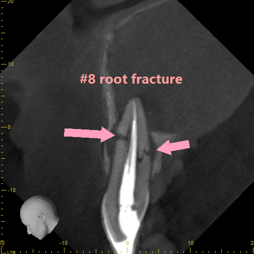 A Sagittal Slice from A CBCT Volume Shows a Root Fracture
Clinical Benefits and Diagnostic Advantages
The integration of CBCT technology into dental practice delivers multiple clinical benefits that translate directly into improved patient care and treatment outcomes. Enhanced diagnostic accuracy represents perhaps the most significant advantage, as the three-dimensional perspective reveals anatomical details and pathological conditions that remain hidden on traditional 2D radiographs.
Improved treatment outcomes result from the precise planning capabilities that cone beam CT imaging provides. Whether planning dental implants, orthodontic movement, or surgical procedures, the detailed anatomical information available through CBCT scans enables clinicians to execute treatments with greater precision and predictability.
Patient communication and case presentation benefit tremendously from the visual impact of 3D imaging. Patients readily understand treatment recommendations when presented with precise, three-dimensional representations of their anatomical conditions. This enhanced communication typically leads to improved case acceptance and patient compliance.
Comprehensive documentation provided by CBCT imaging creates detailed baseline records that support treatment monitoring and legal protection. The ability to measure anatomical structures precisely and document conditions thoroughly enhances the overall quality of patient records.
Time efficiency improvements result from the rapid scanning capabilities and immediate image availability. A single CBCT scan can provide multiple diagnostic views that would otherwise require several traditional radiographic exposures, streamline the diagnostic process and reducing patient appointment time.
CBCT Machine Types and Features
Compact CBCT Units
Compact CBCT machines are specifically designed for practices with limited space or focused imaging needs. These space-efficient units typically offer field of view options ranging from 4x4 to 8x8 cm, making them ideal for endodontic applications and single-tooth implant planning.
The lower initial investment cost of compact systems makes CBCT technology accessible to smaller practices and specialists who primarily need focused imaging capabilities. Despite their size limitations, these units maintain the high-resolution imaging quality essential for detailed diagnostic work.
Compact CBCT scanners excel in applications requiring precise visualization of small anatomical areas. Endodontists particularly benefit from the exceptional detail these systems provide for root canal morphology assessment and periapical lesion evaluation.
Installation requirements for compact units are minimal, typically requiring only 8x10 feet of floor space and standard electrical connections. This accessibility allows practices to integrate CBCT imaging without major facility modifications.
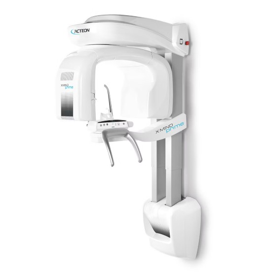 Acteon X-Mind Compact CBCT Unit
Mid-Range CBCT Systems
Mid-range CBCT systems offer the versatility that many general dental practices require, providing adjustable field of view options typically ranging from 4x4cm to 8x10cm. This flexibility allows practitioners to select the optimal scan volume for each clinical situation, balancing diagnostic needs with radiation exposure considerations.
Multiple imaging protocols accommodate various procedures, from focused endodontic evaluation to comprehensive implant planning. The ability to customize scan parameters ensures optimal image quality while minimizing radiation exposure for each examination.
These systems strike an excellent balance between functionality and practice space requirements, making them suitable for general dentistry practices that handle diverse cases and specialist referrals. The moderate investment cost provides access to advanced imaging capabilities without the expense of full-featured systems.
Software packages included with mid-range systems typically offer comprehensive analysis tools for implant planning, orthodontic assessment, and surgical planning applications, supporting the diverse needs of modern dental practice.
 HDXWill EcoX Mid FOV
Full-Featured CBCT Scanners
Full-featured CBCT machines represent the pinnacle of dental imaging technology, offering a large field of view capabilities up to 23x17 cm and comprehensive imaging options that extend beyond basic cone beam CT. These systems often incorporate panoramic and cephalometric imaging modes, providing complete imaging solutions for multi-specialty practices.
Advanced software packages included with full-featured systems offer sophisticated analysis tools for complex treatment planning scenarios. Features such as automatic nerve tracing, airway analysis, and comprehensive implant planning modules support the most demanding clinical applications.
Multi-modal imaging capabilities allow these systems to replace multiple pieces of radiographic equipment, consolidating imaging needs into a single, comprehensive platform. This consolidation can improve workflow efficiency and reduce equipment maintenance costs.
These systems are particularly well-suited for oral surgery centers, orthodontic practices, and multi-specialty clinics that require maximum flexibility and capability to meet diverse imaging needs. Investing in full-featured systems typically yields the best return in high-volume practices with varied case complexity.
Leading CBCT Manufacturers and Models
The CBCT scanner market is characterized by several established manufacturers, each offering distinct advantages and specialized features tailored to meet diverse practice needs.
J.Morita is the first company to introduce Dental CBCT to the US market. J.Morita’s CBCTs are known for image quality and flexibility. JMorita’s X-800 line has FOVs perfect for procedures from endodontics to oral surgery and orthodontics.
Carestream’s CS Series includes the versatile 8100 3D and comprehensive 9600 models, designed to integrate seamlessly with digital practice workflows. The CS 3D Imaging Software provides intuitive tools for implant planning, orthodontic analysis, and thorough treatment planning applications.
HDXWill’s EcoX and DentriMax epitomize excellence when it comes to MAR algorithms and an ever-evolving reconstruction algorithm
Vatech’s Green CT Series combines compact design with AI-enhanced imaging features, offering models suitable for focused applications through comprehensive oral and maxillofacial imaging. Their proprietary EZVIEW software simplifies image analysis and treatment planning workflows.
Radiation Safety and Dose Optimization
Modern CBCT machines incorporate sophisticated dose optimization features that significantly reduce radiation exposure compared to conventional medical imaging. Ultra-low-dose protocols available in current systems deliver effective doses ranging from 11 to 674 micro sieverts, depending on the imaging area and selected parameters.
The radiation dose comparison with traditional medical CT scans reveals the safety advantages of dental cone beam ct. Typical CBCT imaging delivers 200-300 times less radiation exposure than conventional CT scans of equivalent anatomical areas, making the technology suitable for routine diagnostic applications.
ALARA principles (As Low As Reasonably Achievable) guide the design and operation of modern CBCT systems. Automatic exposure control features adjust radiation output based on patient size and density, ensuring optimal image quality with minimal radiation exposure.
Patient-specific protocols allow practitioners to customize scan parameters based on diagnostic requirements and patient characteristics. Pediatric protocols, for example, employ reduced radiation settings while maintaining diagnostic quality appropriate for smaller anatomical structures.
Safety features integrated into modern CBCT machines include movement artifact correction, which automatically adjusts for minor patient movement during scanning, and collision detection systems that prevent equipment contact with patients during rotation.
CBCT Investment Considerations
The financial investment in CBCT technology varies significantly based on system capabilities and features, with initial equipment costs ranging from $50,000 for compact units to $100,000+ for full-featured systems with comprehensive software packages.
Return on investment calculations for CBCT machines typically demonstrate payback periods of 12-24 months in practices with appropriate utilization levels. The revenue generation potential stems from expanded service offerings, reduced referral costs, and improved treatment acceptance rates that result from enhanced diagnostic capabilities.
New service offerings enabled by cbct imaging often include implant planning consultations, advanced endodontic treatments, and comprehensive orthodontic evaluations. These services typically command premium fees while providing significant value to patients through improved treatment outcomes.
Space requirements for CBCT installation must be carefully considered, with most compact units requiring a minimum of 5x5 feet of floor space and larger systems with a cephalometric attachment needing 7x7 feet or more. Dedicated electrical circuit, and lead shielding may require facility modifications that add to implementation costs.
Training and support considerations include comprehensive staff education on equipment operation, radiation safety protocols, and image interpretation fundamentals. Most manufacturers provide initial training packages, but ongoing education is essential for maximizing the diagnostic potential of CBCT imaging.
Integration with Digital Dental Workflows
Modern CBCT systems are designed for seamless integration with comprehensive digital dental workflows, supporting DICOM compatibility that enables direct communication with practice management software and CAD/CAM systems.
The connectivity features of contemporary CBCT machines facilitate direct data transfer for surgical guide fabrication, enabling guided implant placement and other precision procedures. This integration streamlines treatment planning and execution while improving accuracy and predictability.
Cloud-based solutions are increasingly supporting remote access and specialist consultation capabilities, allowing practitioners to securely share CBCT images with referring doctors and specialists for collaborative treatment planning. This connectivity expands access to expertise while maintaining efficient patient care workflows.
AI-enhanced analysis features are beginning to appear in CBCT software, offering automated measurements and diagnostic assistance that reduce interpretation time while improving consistency. These features support less experienced practitioners while enhancing efficiency for all users.
Patient portal integration allows secure image sharing and treatment communication, enabling patients to access their CBCT images and treatment plans remotely. This transparency enhances patient engagement and understanding while supporting informed consent processes.
The digital workflow integration extends to treatment monitoring and outcome assessment, with CBCT imaging providing objective measures of treatment progress and long-term stability. This capability supports evidence-based practice and continuous quality improvement initiatives.
Modern CBCT technology represents a transformative investment for dental practices committed to providing the highest level of patient care. The combination of enhanced diagnostic capabilities, improved treatment outcomes, and comprehensive integration possibilities makes these systems essential tools for contemporary dental practice.
The selection of appropriate CBCT equipment requires careful consideration of practice needs, patient demographics, and growth objectives. Partnering with experienced imaging specialists ensures optimal system selection and implementation, while providing ongoing support to maximize the clinical and financial benefits of this remarkable technology.
Frequently Asked Questions
How often should CBCT equipment be calibrated? Some manufacturers recommend annual calibration by certified technicians, with daily quality assurance checks performed by staff to ensure optimal image quality and radiation dose accuracy. Always check manufacturer guidelines for calibration and state guidelines for quality assurance testing.
Can CBCT machines replace all traditional dental X-rays? While CBCT provides superior 3D imaging, traditional 2D radiographs remain cost-effective for routine dental examinations, bitewing caries detection, and simple diagnostic needs where 3D visualization isn’t required.
What training is required for operating CBCT equipment? Operators typically need 2-4 hours of initial training covering radiation safety, positioning techniques, and image acquisition protocols, plus ongoing continuing education to maintain certification in some jurisdictions.
How do I determine the right field of view for my practice? Consider your primary applications: endodontics requires 4x4-8x8 cm, single implants need 5x5-8x8 cm, full arch work requires 8x8-16x13 cm, and oral surgery may need up to 16x13-23x17 cm field of view.
What are the typical maintenance costs associated with CBCT equipment? Annual service contracts vary depending on the model. Always ask what the ongoing service costs are with the CBCT that you are interested in.
Schedule Your Free Consultation Today
Ready to elevate your dental practice with advanced CBCT technology?
Discover how a state-of-the-art CBCT machine can transform your diagnostic capabilities and treatment outcomes.
Contact us now to schedule your free consultation and get expert guidance tailored to your specific needs.