
J. Morita
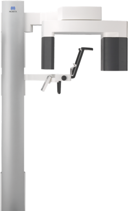
Veraview (X800) R100
Multifunctional J. Morita unit covering both arches, the TMJ, and sinus in a 100×80mm field, at an 80 micrometer voxel size and 2.5 LP/mm resolution.
5.0 · 120 Google Reviews
Serving dentistry since 2003
Installation, training & expert support included
About the Veraview (X800) R100
The Veraview X800 is a newly redesigned multifunctional X-ray unit loaded with practical, unique, and refined features. Image clarity has been enhanced with a voxel size of just 80 micrometers and a resolution of 2.5 LP/mm, the highest level of any Morita multifunctional unit to date. Veraview X800 offers progressive technology that automatically sets the optimal x-ray beam angle, horizontal for 3D or raised 5° for 2D panoramic images. This slight upward projection for panoramic imaging enhances image quality by eliminating potential superimposition of anatomical structures, including the shadow from the base of the skull and hard palate over the maxillary teeth. For 3D imaging, a horizontal beam minimizes distortion and reduces metal artifacts. The X800 unit, therefore, self-adjusts to optimize image quality during both acquisition techniques. The R100 configuration adds the platform's Dental Arch FOV: a Reuleaux-shaped field of view (a rounded triangle, not a cylinder) sized to the curve of the arch itself rather than a circle. At R100 x H80 mm, one scan covers the maxillary and mandibular arches together with the TMJ and maxillary sinus, without the extra dose a cylindrical volume large enough to reach the same corners would require. That makes the R100 the practical mid-tier choice for a practice that regularly needs both arches, sinus, or TMJ in a single acquisition, without stepping up to the F150's larger maxillofacial field. Two dose-management features are built into every X800 configuration, including the R100. Zoom Reconstruction lets you capture a scan at the standard 125 µm voxel size and reconstruct it afterward at the full 80 µm resolution, with no second exposure. Dose Reduction Mode lowers the radiation delivered to lower-density regions of a scan by up to 40% compared to the same scan with it switched off, and a dedicated pediatric panoramic setting narrows the exposure range for smaller jaws. An optional cephalometric arm (R100CP) adds lateral and PA ceph exposures at 100 kV and 96 µm resolution, with an adjustable soft-tissue filter and the option to exclude up to three regions from a partial cephalometric exposure to cut dose further. Every scan, panoramic, CBCT, or ceph, exports in DICOM 3.0 and reads in i-Dixel, with i-Dixel WEB available for browser-based viewing on any PC or tablet without installing dedicated software. The unit's face-to-face design lets the operator position the patient from the front using laser-beam alignment, with a control panel that swings to either side and a chinrest that lowers to about 34 inches for wheelchair access. The X800 platform won an iF Gold Award in 2017 for that design work.
- FOVs: Ø 40 x H 40 mm, Ø 40 x H 80 mm, Ø 80 x H 40 mm, Ø 80 x H 50 mm, Ø 80 x H 80 mm; 3D Reuleaux Full Arch FOVs: Ø 100 x H 40 mm, Ø 100 x H 50 mm, Ø 100 x H 80 mm
- Minimum voxel size: 80 µm
- Resolution: 2.5 LP/mm
- Automatic beam-angle optimization: horizontal for 3D, raised 5° for 2D panoramic
- Focal spot: 0.5mm
- Software: i-Dixel, Exoplan (optional)
- Space requirements: 55.12" x 47.24" (Width x Depth)
- 3 Year Manufacturer Warranty on Parts
- Scan modes: 360° for full-detail acquisition (≈17.9 sec) or 180° for a faster, lower-dose scan (≈9.4 sec)
- Dose Reduction Mode: lowers radiation in lower-density regions of a scan by up to 40%
- Zoom Reconstruction: reconstructs an 80 µm image from a 125 µm capture with no second exposure
- Dental Arch (3D Reuleaux) FOV: arch-shaped rather than cylindrical volume, covering both arches, TMJ, and sinus in one scan
- Panoramic Scout and two-direction scout positioning for accurate, low-stress limited-field CBCT targeting
- Dedicated pediatric panoramic setting with a narrower exposure range for smaller jaws
- Panoramic exposure options: standard, orthographic, and shadowless panoramic; pediatric panoramic; maxillary sinus (anterior/posterior); quadruple TMJ; partial panoramic; bite-wing
- Optional cephalometric arm (R100CP): 100 kV, 96 µm resolution, adjustable soft-tissue filter, partial cephalometric with up to 3 excludable regions
- Face-to-face positioning with a swiveling control panel and a chinrest that lowers to approximately 34" (865 mm) for wheelchair access
- i-Dixel WEB browser-based viewing on any PC or tablet, no dedicated software install required
- DICOM 3.0 compliant: modality worklist management, storage, modality performed procedure step, and print management service classes
- iF Gold Award (2017) for product design
New to CBCT, or narrowing down field of view, voxel size, and cost? Read the complete guide to dental CBCT before you decide.
Under the hood
Veraview (X800) R100 technology
Adaptive Focal Point (AFP)
AFP builds the panoramic image from multiple image-layer captures and blends them so the whole arch, from root apex to incisal edge, stays in focus rather than only the layer matched to an assumed average jaw shape. In practice that means fewer retaken panoramics when a patient's arch curvature falls outside the standard focal trough.
Adaptive Gray Scale (AGS)
AGS reads the density differences across a panoramic image and adjusts contrast automatically, so the dental arch, jaw bone, and TMJ are all clearly readable in the same image instead of forcing a choice between exposing for bone density or for tooth structure. That saves a retake when a case spans both dense and thin anatomy in one shot.
Digital Direct Auto Exposure (DDAE)
DDAE has the flat panel detector read X-ray transparency in real time during the exposure itself and adjusts tube output on the fly, rather than fixing settings beforehand and hoping they hold across the whole scan. The result is a wider dynamic range in a single acquisition, useful when a patient's anatomy varies significantly across the field.
Zoom Reconstruction
Zoom Reconstruction captures a scan at the standard 125 µm voxel size, then reconstructs that same raw data at the finer 80 µm resolution afterward, with no second exposure. If a case needs a closer look once you're already reviewing the images, you get the extra detail without bringing the patient back in for another scan.
3D Reuleaux FOV
Instead of a cylindrical scan volume, the R100 uses a Reuleaux (rounded-triangle) shaped field tailored to the shape of a dental arch. Because the volume doesn't waste dose imaging the empty space a circular FOV would include past the arch's curve, a full-arch scan on the R100 delivers less radiation than a cylindrical FOV large enough to cover the same arch would require.
Dose Reduction Mode
Dose Reduction Mode lowers X-ray output specifically in the more radiolucent regions of a scan, where less radiation is needed to produce a readable image, cutting overall patient dose by as much as 40% compared to the same scan with the mode off. It's one of several dose-control features on the X800 platform, alongside 180°/360° mode selection and the dedicated pediatric panoramic setting, that support following the ALARA principle without giving up diagnostic image quality.
See it in action
Veraview (X800) R100 on video
X800 R100 Perfect Installation
1:27Newest Generation vs. Previous Generation Scan Review
9:13More walkthroughs and training clips in thevideo library.
Veraview (X800) R100 questions, answered
What field of view does the J. Morita Veraview X800 R100 have?
The J. Morita Veraview X800 R100 offers fields of view from Ø 40 x H 40 mm up to Ø 100 x H 80 mm, including 3D Reuleaux Full Arch options at Ø 100 x H 40 mm, Ø 100 x H 50 mm, and Ø 100 x H 80 mm.
What is the resolution of the Veraview X800 R100?
The Veraview X800 R100 has a minimum voxel size of 80 µm and a resolution of 2.5 LP/mm, the highest level of any Morita multifunctional unit to date, with a 0.5 mm focal spot.
How does the Veraview X800 R100 optimize image quality for panoramic versus 3D imaging?
The Veraview X800 R100 automatically sets the optimal beam angle: horizontal for 3D to minimize distortion and reduce metal artifacts, and raised 5° for 2D panoramic images to eliminate superimposition of structures such as the base of the skull and hard palate over the maxillary teeth.
What software does the Veraview X800 R100 work with?
The Veraview X800 R100 works with i-Dixel and optional Exoplan software. Every scan also exports in the standard DICOM format, so it's compatible with any DICOM-compliant viewing or planning software your practice already uses, and Dental TI integrates it as part of installation.
What is included when I buy the Veraview X800 R100 from Dental TI?
Every Veraview X800 R100 purchase from Dental TI includes site planning and installation, integration with your practice's imaging software, on-site team training (remote also available), and expert remote and on-site support. Financing is available, and the unit carries a 3 year manufacturer warranty on parts.
How much space does the Veraview X800 R100 require?
The Veraview X800 R100 requires a footprint of 55.12 inches wide by 47.24 inches deep, and Dental TI handles site planning before installation.
What does 3D Reuleaux FOV mean, and why does the shape matter?
3D Reuleaux FOV is Morita's term for a scan volume shaped like a rounded triangle (a Reuleaux shape) instead of a cylinder, sized to match the curve of a dental arch. Because it doesn't image the empty space outside the arch that a circular field of the same width would include, it captures a full arch at a lower dose than a cylindrical FOV large enough to cover the same area.
Can the Veraview X800 R100 image both arches in one scan?
Yes. The R100's largest field of view, R 100 x H 80 mm, uses the Reuleaux-shaped Dental Arch FOV to capture the maxillary and mandibular arches together with the TMJ and maxillary sinus in a single acquisition, which is the R100's core use case.
What's the difference between the R100 and F150 field-of-view tiers?
The R100 and F150 are the same Veraview X800 hardware configured for different maximum fields of view. The R100 tops out at R 100 x H 80 mm, the Reuleaux-shaped Dental Arch FOV covering both arches, the TMJ, and the sinus. The F150 adds a larger Ø 150 x H 140 mm maxillofacial field on top of everything the R100 offers, useful for orthodontic, full-jaw, or craniofacial cases that need more than an arch-level view. The F40, by contrast, is limited to Ø 40 mm FOVs for single-site endodontic and implant work.
How much can the Veraview X800 R100 reduce a patient's X-ray dose?
Several dose-control features work together on the R100: Dose Reduction Mode lowers radiation in lower-density regions of a scan by up to 40% compared to the same scan with it off, the 180° scan mode trades some detail for a faster, lower-dose acquisition than the 360° mode, and a dedicated pediatric panoramic setting narrows the exposure range for smaller jaws. Which combination makes sense depends on the case.
Does the Veraview X800 R100 support cephalometric imaging?
Yes, as the R100CP configuration, which adds a cephalometric arm to the same panoramic/CBCT unit. It captures lateral and PA cephalometric exposures at 100 kV and 96 µm resolution, with an adjustable soft-tissue filter and the option to exclude up to three regions from the exposure to reduce dose further.
Is the Veraview (X800) R100 right for your practice?
Talk it through with an advisor who installs these machines every week — including the ones we'd steer you away from.