
PreXion
Excelsior Mid (MFOV)
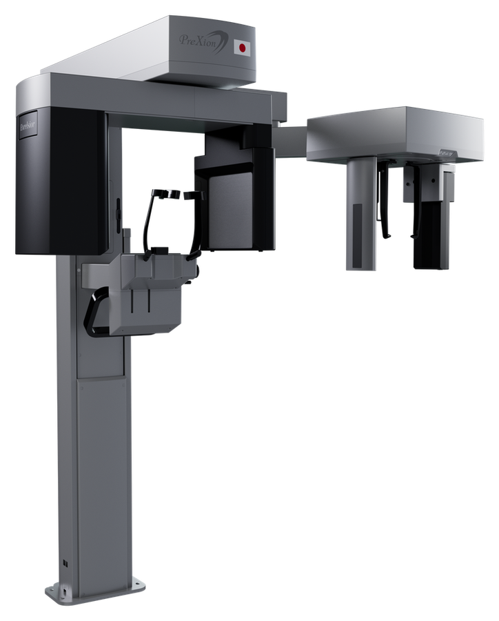
PreXion's medium field of view CBCT — full-arch coverage in one scan, with the same PMC and MAR imaging suite as the rest of the current Excelsior line.
Serving dentistry since 2003
Installation, training & expert support included
About the Excelsior Mid (MFOV)
The Excelsior Mid (MFOV) is PreXion's medium field-of-view CBCT: a combination panoramic and 3D cone-beam unit that steps up from single-arch, small-FOV scanning to a full 16×9 cm field, covering an entire arch — mandible, maxilla, airways, sinuses, and TMJ — in a single volume. The same unit still switches down to 9×9 cm (complete mandible), 9×6 cm (single arch and TMJ), and 5×5 cm (implant, endo, and 3rd-molar detail) for lower-dose, targeted exams, so a practice isn't locked into a large-volume exposure for every case. In PreXion's current Excelsior lineup, the Mid sits between the Excelsior Endo and the Excelsior Max. The Endo trades field-of-view range (up to 14×9 cm) for a smaller 0.2mm focal spot and voxels as fine as 48 µm, built specifically for endodontic detail. The Max keeps the Mid's same small-FOV options but extends up through 16×15 and 16×21 cm for full craniofacial and orthodontic cases. The Excelsior Mid is the middle ground: full-arch coverage for implant planning, general restorative work, and orthodontic case review, without paying for cranial-scale field of view a general practice may never use. Both upgrade paths stay open after purchase. PreXion documents a field upgrade from Excelsior Mid to Excelsior Mid CEPH (adding cephalometric capture to the installed unit) and a separate field upgrade from Excelsior Mid to Excelsior Max (extending the field of view), so a practice that outgrows the Mid's 16×9 cm ceiling isn't stuck buying a whole new system. It shares the current Excelsior platform's imaging pipeline: Patient Motion Correction (PMC), 3-level Metal Artifact Reduction (MAR), 120 kV tube operation, and Software Eye for acquisition, viewing, and DICOM workflow, with 3D implant planning, airway volume measurement, STL conversion, virtual endoscopy, and DICOM fusion built in, plus AI-assisted cephalometric tracing once the Ceph option is added. The same unit also runs a full 2D panoramic suite — standard, pediatric, and fast panoramic modes, bitewing, TMJ, maxillary sinuses, enhanced ortho, and 41-layer multi-slice panoramic — with its own contrast and focus algorithms for image quality, plus a 3-axis movement system (two orthogonal directions and one rotation) for flexible radiographic profiles.
- FOVs: 5×5 cm (implant/endo/3rd-molar detail), 9×6 cm (single arch + TMJ), 9×9 cm (full arch), 16×9 cm (extended mandible, airways, sinuses, TMJ)
- Voxel size: 75–400 µm, isotropic
- Focal spot: 0.5mm
- Tube: 60–120 kV, 3.2–16 mA — 120 kV operation reduces beam-hardening artifacts and low-energy photon noise
- Resolution programs: LD, STD, HD, and UHD — choose exposure time and voxel size per exam, trading resolution against dose
- Patient Motion Correction (PMC) and 3-level Metal Artifact Reduction (MAR) — MAR levels can be reprocessed after the scan without re-exposing the patient
- Software Eye includes 3D implant planning, airway volume measurement, STL conversion, virtual endoscopy, and DICOM fusion; AI-assisted cephalometric tracing is available once the Ceph option is added
- Full 2D panoramic suite: standard, pediatric, and fast panoramic modes, bitewing, TMJ, maxillary sinuses, enhanced ortho, and 41-layer multi-slice panoramic, with dedicated contrast and focus algorithms
- 3-axis movement system (two orthogonal directions plus rotation) for flexible radiographic profiles and constant vertical magnification
- Field-upgradeable: add cephalometric imaging (Excelsior Mid CEPH), or upgrade to the larger Excelsior Max field of view
- Software: Software Eye (1 server + 5 clients license)
- Supply voltage: 110/127/220/240 AC
New to CBCT, or narrowing down field of view, voxel size, and cost? Read the complete guide to dental CBCT before you decide.
Under the hood
Excelsior Mid (MFOV) technology
Patient Motion Correction (PMC)
Small patient movements during a CBCT exposure can blur the reconstructed volume enough to force a retake. PMC automatically corrects for these micro-movements in the captured data, so exams come out at diagnostic quality the first time. Fewer retakes means less dose spent on repeat scans and less time lost rescheduling a patient.
Metal Artifact Reduction (MAR)
Implants, posts, gutta-percha, and wide restorations create bright-metal artifacts that can obscure the anatomy around them. The Excelsior Mid corrects for this with three processing levels plus automatic metal reduction, and because the correction runs on the reconstructed volume, you can reprocess an existing scan at a different level whenever a case calls for it, without bringing the patient back for another exposure.
120 kV Tube Voltage
The Excelsior Mid's tube runs up to 120 kV, paired with radiation filters tuned for a higher average beam energy. That combination cuts down on beam-hardening artifacts and reduces the low-energy photon noise that would otherwise soften fine detail, so images come out cleaner without an extra post-processing step.
3D Implant Planning
Software Eye's implant planning tool lets you simulate implant position directly on the 2D and 3D reconstruction, identify the mandibular canal, and take the measurements you need before you're in the surgical field. It runs on the same acquisition PC used for the scan, so there's no separate license or workstation to manage for planning a case.
Airway Volume Measurement
The Excelsior Mid's 16×9 cm field of view captures the mandible, maxilla, airways, sinuses, and TMJ in one volume, and Software Eye includes an airway volume measurement tool to quantify that anatomy directly from the scan — useful for sleep-related airway evaluation without ordering a separate exam.
AI-Assisted Cephalometric Tracing
Adding the Ceph option to an Excelsior Mid (Excelsior Mid CEPH) brings cephalometric capture and manual tracing across protocols including McNamara, USP, Ricketts, Steiner, Rocabado, and Tweed. An optional AI-assisted tracing module can apply those same recognized protocols automatically, cutting down the time a complex analysis takes.
Sample captures
Clinical output from the Excelsior Mid (MFOV)
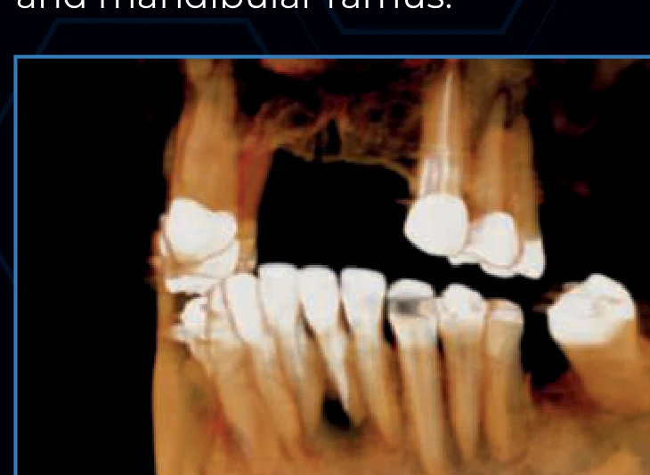
Actual radiographs produced by this system — the image quality you're buying, not a brochure rendering.
Excelsior Mid (MFOV) questions, answered
What field of view options does the PreXion Excelsior Mid have?
The Excelsior Mid (MFOV) offers four fields of view: 5×5 cm, 9×6 cm, 9×9 cm, and 16×9 cm — the 16×9 cm option covers a full arch in a single scan.
What is the difference between the Excelsior Mid and Excelsior Max?
Both cap out at a 16 cm diameter field of view, but the Excelsior Max extends taller — up to 16×21 cm versus the Mid's 16×9 cm — for cases that need cranial or extended full-arch coverage.
Can the PreXion Excelsior Mid capture cephalometric images?
Yes, as an option — the Excelsior Mid CEPH configuration adds cephalometric capture to the same unit.
What is the voxel size and focal spot of the PreXion Excelsior Mid?
The Excelsior Mid has a focal spot of 0.5mm and an isotropic voxel size from 75 to 400 µm depending on the field of view and resolution selected.
What software does the PreXion Excelsior Mid work with?
The Excelsior Mid runs Software Eye with PMC motion correction and 3-level MAR. Every scan also exports in the standard DICOM format, so it's compatible with any DICOM-compliant viewing or planning software your practice already uses, not just Software Eye.
What is included when I buy the PreXion Excelsior Mid from Dental TI?
Every PreXion Excelsior Mid comes with manufacturer-backed installation and support from PreXion. Dental TI adds another layer on top: site planning, integration with your practice's imaging software, on-site team training with remote training also available, and expert remote and on-site support of our own. Dental TI has served dentistry since 2003 from Carmel, Indiana.
Can the Excelsior Mid be upgraded to a larger field of view or cephalometric imaging later?
Yes. PreXion documents two upgrade paths from the Excelsior Mid: adding cephalometric capture to the same unit (Excelsior Mid → Excelsior Mid CEPH), or upgrading the field of view to the Excelsior Max (Excelsior Mid → Excelsior Max). Both are field upgrades to your installed unit, not a trade-in.
Does correcting metal artifacts on the Excelsior Mid require rescanning the patient?
No. The Excelsior Mid's 3-level Metal Artifact Reduction lets you reprocess the same scan at a different correction level after the fact — for gutta-percha, implants, wide prostheses, or metal restorations — without a new exposure.
What computer do I need to run the Excelsior Mid?
PreXion's minimum recommendation for the Excelsior Mid's 3D acquisition workstation: Windows 10 64-bit Professional, Intel Core i7 (10th gen or newer, 12 MB cache, 4.0 GHz+), 16 GB RAM, an Nvidia GeForce RTX 4060 8GB or better, a 500W+ power supply, 1TB+ storage, a 1920×1080 monitor, and a dedicated Gigabit Ethernet adapter.
Does installing the Excelsior Mid require a certified technician?
Yes — PreXion requires equipment assembly to be performed by a certified technician, and the shipping packaging must stay sealed until they arrive; opening it early voids the manufacturer's warranty. Dental TI coordinates this as part of installation.
Does the Excelsior Mid include implant planning software?
Yes — 3D implant planning is standard in Software Eye on every Excelsior Mid: you can simulate implant position in 2D and 3D, identify the mandibular canal, and take measurements directly on the acquisition PC before you're in the surgical field.
Does the Excelsior Mid support airway or sinus evaluation?
Yes. The 16×9 cm field of view captures the mandible, maxilla, airways, sinuses, and TMJ in a single volume, and Software Eye includes an airway volume measurement tool to quantify that anatomy from the same scan.
What cephalometric analysis protocols does the Excelsior Mid CEPH support?
Manual tracing supports named protocols including McNamara, USP, Ricketts, Steiner, Rocabado, and Tweed. An optional AI-assisted tracing module can also apply these scientifically recognized protocols automatically to speed up complex analyses.
What is the difference between the Excelsior Mid and Excelsior Endo?
The Excelsior Endo trades field-of-view range for resolution: it caps at 14×9 cm (versus the Mid's 16×9 cm) but uses a smaller 0.2mm focal spot and resolves down to a 48 µm voxel, versus the Mid's 0.5mm focal spot and 75 µm minimum voxel. The Endo also has no cephalometric option. Choose the Mid for full-arch coverage with a cephalometric upgrade path; choose the Endo when endodontic image detail is the priority.
Can the Excelsior Mid also take panoramic and bitewing images?
Yes — the Excelsior Mid is a combination panoramic and 3D CBCT unit. It includes standard, pediatric, and fast panoramic modes, bitewing, TMJ, maxillary sinuses, enhanced orthodontic, and a 41-layer multi-slice panoramic mode, all in the same Software Eye acquisition software as the 3D scans.
Is the Excelsior Mid (MFOV) right for your practice?
Talk it through with an advisor who installs these machines every week — including the ones we'd steer you away from.