
J. Morita
Veraview (X800) F40
Multifunctional J. Morita unit with a focused 40×80mm field for endodontic and single-site detail, at an 80 micrometer voxel size and 2.5 LP/mm resolution.
Serving dentistry since 2003
Installation, training & expert support included
About the Veraview (X800) F40
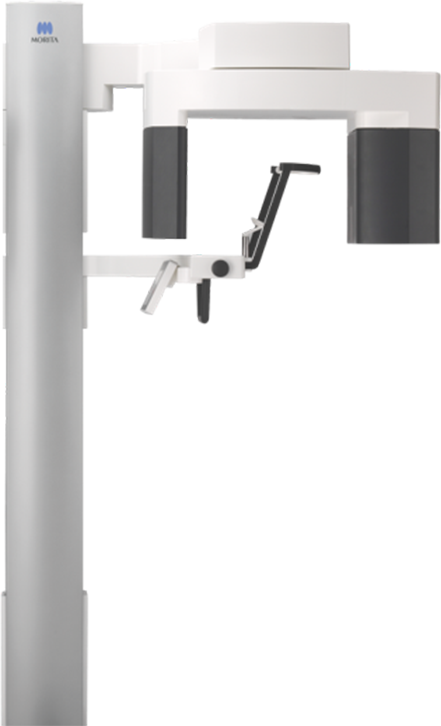
The Veraview X800 is J. Morita's current hybrid imaging platform: one chassis, one imaging engine, sold across a handful of field-of-view tiers that Dental TI names by scan volume — F40, R100, and F150. The F40 is the smallest and most focused of the three. It ships with exactly two fields of view, Ø 40 x H 40 mm and Ø 40 x H 80 mm, both scoped to a single tooth or a short span of adjacent teeth rather than the full arch. That narrow scope is the point of the tier: endodontic diagnosis, single-implant planning, and root or canal detail work don't need a wider volume, and a smaller volume means a faster reconstruction and less dose to the patient than the platform's R100 or F150 configurations, which are built for full-arch and orthodontic work instead. Resolution is where the F40 does its real work. At 80 micrometer minimum voxel size and 2.5 LP/mm at 10% MTF, it's the highest resolution of any Morita multifunctional unit to date, sharp enough to separate canal anatomy, periapical bone changes, and root fracture lines that a coarser 125 micrometer scan would blur together. Zoom Reconstruction lets a clinician capture at the faster, lower-dose 125 micrometer setting by default and reprocess that same dataset up to 80 micrometers afterward, with no second exposure, so the extra detail is available on demand rather than something the practice has to commit to on every scan. For CBCT exposures the X800 scans with a horizontal x-ray beam, which minimizes distortion and metal artifact compared to the raised-beam geometry used for panoramic capture; the unit shifts its Flat Panel Detector to raise that beam 5° automatically when it switches to panoramic mode, which keeps the hard palate and skull base from shadowing the maxillary teeth. A 360°/180° scan-mode toggle trades a slower, more detailed 360° capture against a faster 180° pass at a lower dose, and Dose Reduction Mode further lowers output in the less-dense areas of a given scan by as much as 40% compared with the function switched off. Panoramic Scout lets a clinician frame a limited-field CBCT directly from a panoramic image rather than positioning blind. On the 2D side, Adaptive Focal Point, Adaptive Gray Scale, and Digital Direct Auto Exposure work together to keep a panoramic exposure evenly in focus, evenly dense, and correctly exposed from root apex to incisal edge, whether the case is a routine screening or a detail read alongside a CBCT volume. The unit's face-to-face design — a chinrest that lowers to accommodate a wheelchair, and a control panel that swings to either side of the patient for better sightlines during positioning — won J. Morita an iF Gold Award in 2017 for design and ergonomics, and every exposure is DICOM 3.0-compliant, viewable in i-Dixel or from any browser through i-Dixel WEB.
- FOVs: Ø 40 x H 40 mm, Ø 40 x H 80 mm
- Minimum voxel size: 80 µm
- Resolution: 2.5 LP/mm
- Automatic beam-angle optimization: horizontal for 3D, raised 5° for 2D panoramic
- Focal spot: 0.5mm
- 360°/180° scan mode toggle: 360° for maximum detail, 180° for a faster scan at lower dose
- Zoom Reconstruction: capture at 125 µm, reconstruct the same dataset to 80 µm with no repeat exposure
- Dose Reduction Mode: up to 40% lower radiation in the less-dense areas of a scan
- Panoramic Scout: frame a limited-field CBCT directly from a panoramic image
- AFP, AGS, and DDAE panoramic processing for consistent focus, density, and dynamic range
- Pediatric Panoramic: narrower exposure range sized to children's smaller jaws
- Face-to-face design: chinrest lowers to 34" (865 mm) for wheelchair access, control panel swings to either side of the patient
- i-Dixel WEB: view CBCT and 2D images from any browser, no software install required
- iF Gold Award, 2017, for design and ergonomics
- Software: i-Dixel, Exoplan (optional)
- Space requirements: 55.12" x 47.24" (Width x Depth)
- 3 Year Manufacturer Warranty on Parts
New to CBCT, or narrowing down field of view, voxel size, and cost? Read the complete guide to dental CBCT before you decide.
Under the hood
Veraview (X800) F40 technology
Adaptive Focal Point (AFP)
AFP tiles the panoramic exposure across multiple image layers, from just outside the arch to just inside it, and keeps the reconstruction sharp along whichever layer the case calls for. Because arch depth varies by patient, that per-layer optimization is what keeps a panoramic image evenly in focus from the root apex to the incisal edge, instead of sharp through the molars and soft through the anteriors, or the other way around.
Adaptive Gray Scale (AGS)
AGS adjusts image density automatically across a panoramic exposure, so the dental arch, the denser jaw bone, and the TMJ all read clearly in the same image rather than needing separate exposures or manual contrast windowing afterward. It runs alongside AFP, and the two can be toggled independently to match the read a case needs.
Digital Direct Auto Exposure (DDAE)
The Flat Panel Detector reads x-ray transparency in real time during the exposure itself, and the system adjusts x-ray output on the fly instead of relying on one pre-set exposure value for the whole capture. That in-exposure correction is what gives the image its dynamic range — dense restorations and thin bone both show up in the same read without one blowing out or the other going dark.
Zoom Reconstruction
A standard capture runs at 125 µm voxel size for a lower dose and a faster scan. Zoom Reconstruction reprocesses that same dataset afterward at the X800's 80 µm high-resolution setting, with no second exposure and no added dose to the patient. On the F40's Ø40 fields of view, that means a practice can default to the faster capture and pull the extra detail only when a case turns out to need a closer look at canal anatomy — Morita disables the function only at the platform's largest Ø150 field of view, which the F40 tier doesn't offer in the first place.
3D Reuleaux FOV
On the X800 platform's larger R100 and F150 configurations, the Dental Arch field of view is shaped to the curve of the arch itself — a rounded triangle rather than a cylinder — so a full-arch scan covers the region of interest without irradiating the tissue around it that a plain cylindrical volume would include. The F40's own fields of view (Ø40 x H40 mm and Ø40 x H80 mm) are already scoped to a single site rather than the full arch, but it's the same platform principle at work: shape the scan volume to the target instead of the other way around.
Dose Reduction Mode
Tissue density varies within a single scan, and Dose Reduction Mode lowers x-ray output specifically in the more transparent areas rather than exposing the whole volume at one setting. Morita's own testing shows a reduction of as much as 40% compared with the function switched off. On the F40, that stacks on top of the dose savings already built into scanning a Ø40 field of view instead of a full-arch volume — the same as-low-as-reasonably-achievable approach that runs through the platform, including a narrower Pediatric Panoramic exposure range for children's smaller jaws.
Veraview (X800) F40 questions, answered
What field of view does the J. Morita Veraview X800 F40 have?
The J. Morita Veraview X800 F40 offers two focused fields of view: Ø 40 x H 40 mm and Ø 40 x H 80 mm, keeping the scan volume tight to the region of interest.
What is the resolution of the Veraview X800 F40?
The Veraview X800 F40 has a minimum voxel size of 80 µm and a resolution of 2.5 LP/mm, the highest level of any Morita multifunctional unit to date, with a 0.5 mm focal spot.
How does the Veraview X800 F40 optimize image quality for panoramic versus 3D imaging?
The Veraview X800 F40 automatically sets the optimal beam angle: horizontal for 3D to minimize distortion and reduce metal artifacts, and raised 5° for 2D panoramic images to eliminate superimposition of structures such as the base of the skull and hard palate over the maxillary teeth.
What software does the Veraview X800 F40 work with?
The Veraview X800 F40 works with i-Dixel and optional Exoplan software. Every scan also exports in the standard DICOM format, so it's compatible with any DICOM-compliant viewing or planning software your practice already uses, and Dental TI integrates it as part of installation.
What is included when I buy the Veraview X800 F40 from Dental TI?
Every Veraview X800 F40 purchase from Dental TI includes site planning and installation, integration with your practice's imaging software, on-site team training (remote also available), and expert remote and on-site support. Financing is available, and the unit carries a 3 year manufacturer warranty on parts.
What is Adaptive Focal Point (AFP) and how does it improve panoramic image sharpness?
AFP tiles a panoramic exposure across multiple image layers and reconstructs each one to stay sharp on its own, so the final image reads evenly in focus from the root apex to the incisal edge instead of sharp in one region and soft in another. It works alongside Adaptive Gray Scale (AGS) for density and Digital Direct Auto Exposure (DDAE) for dynamic range, and each can be toggled independently.
How much can Dose Reduction Mode lower radiation exposure on the Veraview X800?
Dose Reduction Mode lowers x-ray output in the more transparent areas of a scan rather than exposing the entire volume at one setting, and Morita's testing shows a reduction of as much as 40% compared with the function switched off. On the F40, that's on top of the dose savings that already come from scanning a tight Ø40 field of view instead of a full-arch volume.
Is the Veraview X800 F40 a good fit for endodontic work?
Yes — a focused field of view is the point of the F40 tier. Its Ø 40 x H 40 mm and Ø 40 x H 80 mm fields of view scope a scan to a single tooth or a short span of adjacent teeth rather than the full arch, at 80 µm minimum voxel size and 2.5 LP/mm resolution, sharp enough to separate canal anatomy, periapical bone changes, and root fracture lines. That's a narrower, faster, lower-dose exposure than Dental TI's R100 or F150 configurations of the same X800 platform, which are built for full-arch and orthodontic work instead.
Does the Veraview X800 F40 support Zoom Reconstruction?
Yes. Zoom Reconstruction reprocesses a standard 125 µm capture up to the platform's 80 µm high-resolution setting without a second exposure. Morita disables the function only at the platform's largest Ø150 field of view, which the F40 tier doesn't offer — both of the F40's own fields of view support it, so a practice can default to the faster, lower-dose capture and pull the extra detail after the fact only when a case needs it.
Can I choose between a 360° and 180° scan on the Veraview X800 F40?
Yes. The unit toggles between a 360° scan (about 17.9 seconds) for maximum detail and a faster 180° scan (about 9.4 seconds) at a lower dose, so the exposure can be matched to what the case actually calls for.
Is the Veraview (X800) F40 right for your practice?
Talk it through with an advisor who installs these machines every week — including the ones we'd steer you away from.