Intraoral Camera for Patient Education Works
- Apr 29
- 6 min read
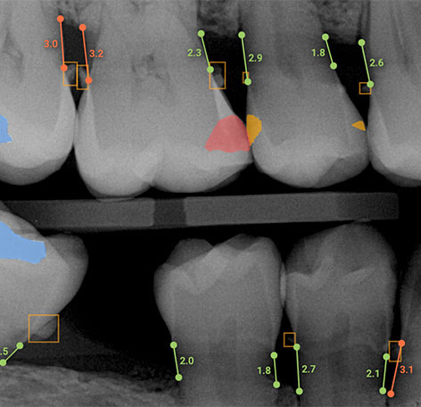
A patient looks at a monitor, sees a crack running across a molar, and the conversation changes immediately. What felt abstract a minute earlier becomes visible and personal. That is why an intraoral camera for patient education has become more than a convenience in modern dentistry - it is a practical communication tool that helps patients understand conditions, recommended treatment, and timing.
For many practices, the challenge is not identifying clinical issues. It is helping patients see why those issues matter now, not six months from now. Verbal explanations alone often leave too much room for uncertainty. Patients may nod during the exam and still leave unconvinced, confused about urgency, or hesitant to move forward. When the clinical team can show a worn margin, fractured cusp, inflamed tissue, or plaque accumulation in real time, education becomes clearer and treatment discussions become more productive.
Why an intraoral camera for patient education changes the conversation
An intraoral camera gives patients a view they would never otherwise have of their own oral condition. That direct visual context often reduces the gap between diagnosis and understanding. Instead of asking patients to trust a description of a failing restoration, clinicians can show the restoration on screen and explain what they are seeing in plain language.
That matters because treatment acceptance is rarely driven by information alone. It is driven by confidence. Patients want to know that the recommendation is justified, that the issue is real, and that the practice is focused on their health rather than simply presenting another procedure. Images support that confidence without adding pressure.
This is especially useful in cases that are easy to delay. Small cracks, early wear, leaking restorations, and soft tissue changes may not be painful yet. When patients can see those conditions, the rationale for preventive or restorative treatment becomes easier to understand. The result is often a better clinical discussion and a faster path to informed consent.
Better visuals, better case acceptance
Most practices evaluate technology based on diagnostic value, workflow impact, and return on investment. An intraoral camera touches all three, but its value in patient education is often where practices see the fastest payoff.
When patients understand what is happening, case acceptance tends to improve. That does not mean every image leads to immediate approval. It means the conversation becomes more grounded, and objections become easier to address. A patient who sees recurrent decay around an existing crown is usually asking different questions than a patient who only hears that the crown should be replaced.
The difference is not just visual proof. It is also specificity. High-quality chairside images allow the team to explain exactly what they are monitoring, where deterioration is occurring, and why treatment now may be simpler and less costly than treatment later. For offices focused on both clinical outcomes and production efficiency, that clarity can support stronger scheduling and fewer delayed decisions.
There is also a trust benefit. Patients generally respond well when a practice takes time to educate rather than persuade. Images create a more collaborative tone. The clinician is not simply recommending care. They are reviewing findings with the patient and inviting questions. That small shift can have a meaningful effect on the overall patient experience.
Where intraoral cameras help most in daily practice
The strongest use cases are often the most routine. Restorative dentistry is an obvious fit because fractures, open margins, failing fillings, and wear patterns are highly visual. Hygiene visits are another strong opportunity. Showing plaque retention, calculus buildup, tissue inflammation, or recession can reinforce home care recommendations and support periodontal conversations.
Cosmetic consultations also benefit. Patients considering whitening, veneers, or alignment usually want to understand both the esthetic issue and the likely outcome. Intraoral images provide a strong baseline for that discussion. In specialty settings, the value continues. Endodontic, pediatric, and oral surgery practices can all use chairside images to improve understanding and reduce uncertainty.
What changes from office to office is not whether the camera is useful, but how consistently the team uses it. In some practices, the camera becomes a standard part of every new patient exam and every restorative consult. In others, it gets used only when a doctor remembers to pull it out. The difference in results usually comes down to process, not hardware alone.
Choosing the right intraoral camera for patient education
Not every camera performs the same in a busy clinical environment. Image quality matters, but it is only one part of the decision. For patient education, clarity, ease of capture, display speed, ergonomic design, and software compatibility all affect how often the device will actually be used.
If the image is grainy, poorly lit, or awkward to capture, the educational value drops quickly. Patients need to see the condition clearly, and the team needs to be able to capture that image without slowing the appointment. A camera that integrates well with imaging software and displays quickly in the operatory supports smoother conversations and less friction during exams.
It also helps to think beyond the initial purchase. Training, implementation support, and long-term service matter more than many practices expect. A camera that looks attractive on paper can still underperform if staff are not comfortable using it or if technical issues interrupt workflow. That is one reason many practices work with consultative technology partners rather than treating the purchase as a simple commodity decision.
Implementation matters as much as the device
Practices do not get the full value of imaging technology by installing it and hoping the team adapts. They get value by building repeatable habits around it. For an intraoral camera, that means deciding when images should be captured, who is responsible, how the doctor reviews them, and how they are presented to the patient.
A simple workflow often works best. Hygienists or assistants can capture key images during exams or recare visits, the doctor can review and annotate findings chairside, and the treatment coordinator can reference those images again when discussing next steps. That continuity keeps the message consistent from operatory to front desk.
Training is central here. Teams need to know not just how to operate the camera, but how to use images effectively in conversation. The goal is not to overwhelm patients with technical detail. It is to explain findings clearly, answer questions, and connect the image to a practical treatment recommendation.
This is where experienced support can make a real difference. Dental TI has built its model around helping practices implement imaging technology with training, consultation, and ongoing service so equipment gets used as intended instead of becoming one more underused purchase.
Common mistakes that limit results
One of the most common issues is inconsistent use. If only certain providers use the camera or if it comes out only for larger cases, the practice misses the broader educational impact. Small findings and preventive conversations often produce some of the best patient engagement because they show patients the value of early intervention.
Another issue is overexplaining. Patients do not need a lecture in material science to understand that a restoration is breaking down. They need a clear image, a straightforward explanation, and a reason the recommendation matters. Simplicity usually improves comprehension.
There is also the question of image quality. Poor angulation, fogging, weak lighting, or cluttered screens can weaken the moment. The visual has to be easy to read. If the patient cannot tell what they are looking at, the camera becomes less persuasive and less efficient.
Finally, some offices underestimate the operational side. If images are hard to save, difficult to retrieve, or disconnected from the patient record, the process can feel cumbersome. Good integration helps the camera support workflow instead of interrupting it.
The ROI is broader than case acceptance alone
Case acceptance gets most of the attention, but the return is broader. Better patient education can reduce confusion, strengthen trust, support documentation, and improve communication across the team. It can also help standardize exams by making findings easier to share and review.
There is a financial benefit to that consistency, but there is also a clinical one. Patients who understand their conditions are often more engaged in both treatment and prevention. They are more likely to follow through, ask better questions, and recognize the value of ongoing care.
For practices evaluating imaging technology, that broader impact matters. The best technology investments do not just produce better images. They support better conversations, better workflows, and better use across the entire patient journey.
An intraoral camera will not fix weak communication on its own, and it will not replace sound diagnosis or a well-trained team. What it can do is make your clinical findings visible in a way patients understand immediately. In a practice focused on clarity, efficiency, and stronger treatment discussions, that is not a small advantage - it is a practical step toward more informed care.