
16 × 15 cm in one scan.
Large-volume coverage in a single acquisition, without stitching for this field of view.
Built for the full picture. Engineered for fine detail.
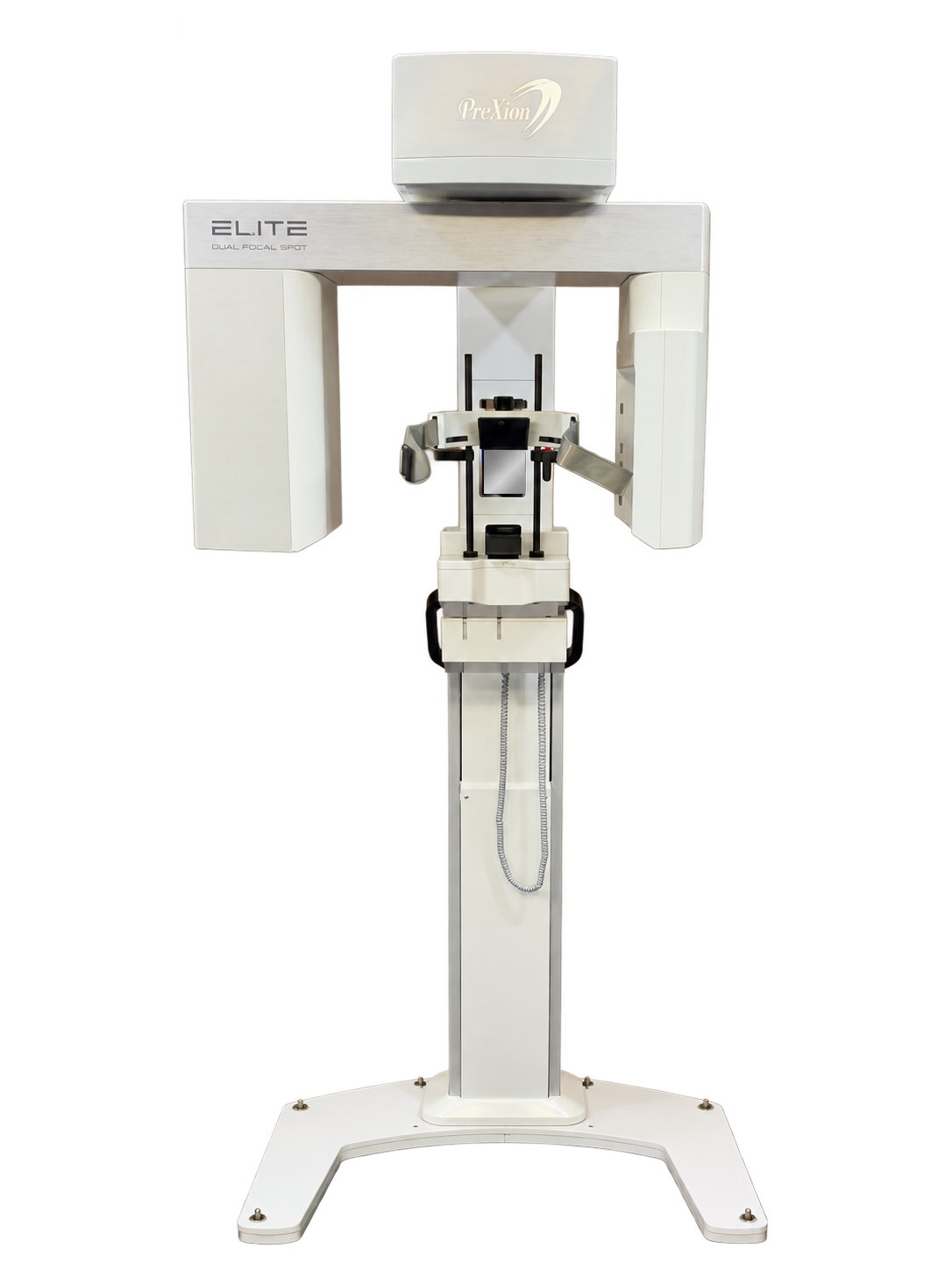
Elite pairs a 0.17 mm CBCT focal spot with voxel sizes down to 48 µm for fine-detail endodontic imaging, plus six fields of view for broader treatment planning. Dedicated panoramic imaging completes the system.
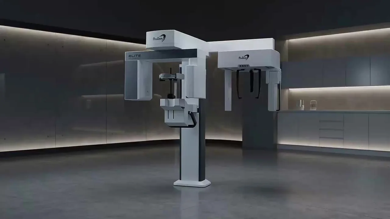
Book an Elite demo Skip to the technologyOptional cephalometric configuration shown.
Choose from six fields of view, from a localized 5 × 5 cm examination to 16 × 21 cm dual-scan coverage. Move through intermediate sizes as the anatomy and clinical question require.
Six fields of view, in centimeters

Large-volume coverage in a single acquisition, without stitching for this field of view.
Use the larger dual-scan field when the examination calls for more coverage, with AI Full Head Stitching.
Match focused endodontic scans or broader treatment-planning views to the anatomy you need to assess.
A 16 × 21 cm full-face study brings skeletal anatomy, a panoramic MIP and an airway view into the same case review.
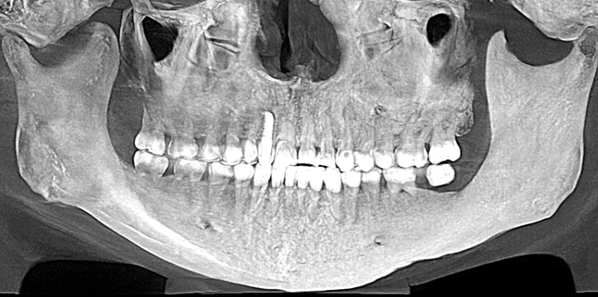
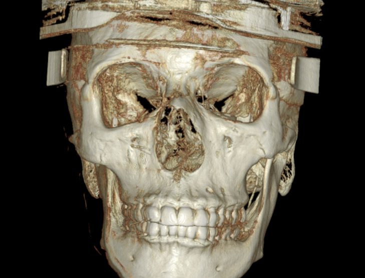
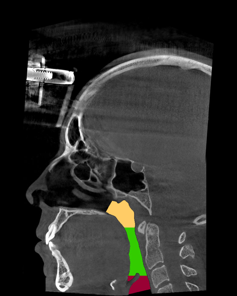
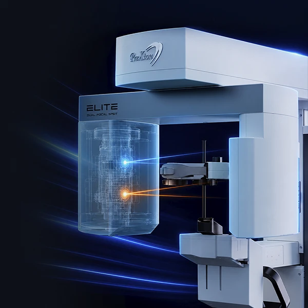
A 0.17 mm focal spot serves 3D CBCT, while panoramic imaging has a dedicated focal spot of its own. Elite brings both imaging modes together with an Ultra HD sensor and a choice of acquisition protocols.
Focal-spot size describes the X-ray source. Voxel size describes the reconstructed 3D volume. They are separate specifications, both part of the complete imaging system.
Fine-detail endodontic protocols offer voxel sizes down to 48 µm. Paired with Elite’s 0.17 mm CBCT focal spot, they give your team a dedicated option for focused endodontic examinations.
The 48–500 µm voxel range lets your team tailor acquisition to the anatomy and examination.
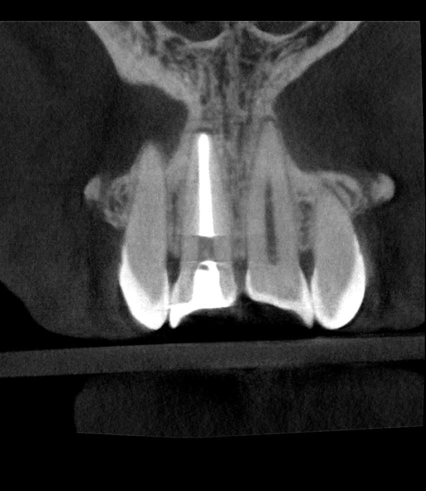
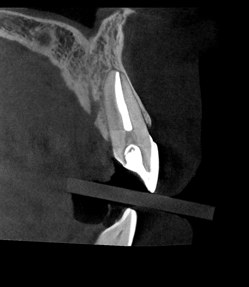
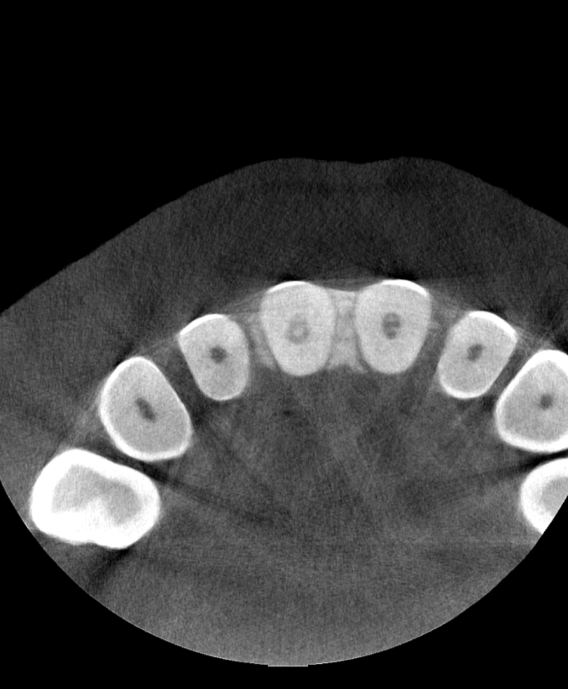
Three views from a 5 × 5 cm endodontic examination. Follow the anterior tooth through coronal, sagittal and axial slices.
Position the patient with fully adjustable APS head support, carbon-fiber arms and multiple stabilization points. Adjust the support to help your team establish and maintain the patient’s position for the scan.
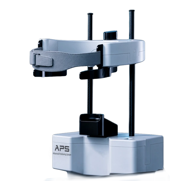
Fully adjustable head support, carbon-fiber arms and multiple stabilization points.
Patient Motion Correction addresses the effects of minor patient movement.
Metal Artifact Reduction helps reduce interference from high-density materials.
Your routine panoramic work belongs alongside your 3D examinations. Elite adds multi-slice review and 2D viewing tools, with a cephalometric configuration available when your practice needs it.
Move through 41 panoramic slices to explore the structures of interest. Optimized 2D viewing filters give your team additional ways to review the image.
Add cephalometric capability to the conversation when your practice needs it. Build the configuration around your imaging work, room and team.
Cephalometric imaging requires the optional configuration. The system and accessories included are specified in your proposal.
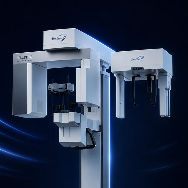
Explore the imaging hardware, acquisition choices, patient support and review tools in one place.
| Imaging modalities | CBCT and dedicated panoramic imaging; optional cephalometric imaging |
|---|---|
| Sensor | Ultra HD sensor |
| Dual focal spots | 0.17 / 0.5 mm |
| CBCT focal spot | 0.17 mm |
| Panoramic focal spot | Dedicated focal spot for PAN |
| Tube voltage | 60–120 kV |
| Cooling | Ultra-fast cooling system |
| All six fields of view | 5 × 5, 9 × 6, 9 × 9, 16 × 9, 16 × 15 and 16 × 21 cm |
|---|---|
| Largest single-scan field | 16 × 15 cmSingle scan |
| Largest dual-scan field | 16 × 21 cmDual scan |
| Voxel range | 48–500 µm |
| Minimum voxel size | 48 µm with high-resolution protocols |
| Extended-volume reconstruction | AI Full Head Stitching |
| Panoramic acquisition | Dedicated PAN imaging |
|---|---|
| Multi-slice panoramic review | 41 panoramic slices |
| 2D image review | Optimized 2D viewing filters |
| Cephalometric imaging | Optional configuration |
| Patient support | APS, Advanced Positioning System |
|---|---|
| Head support | Fully adjustable |
| Support-arm construction | Carbon-fiber arms |
| Stabilization | Multiple stabilization points |
| Patient Motion Correction (PMC) | Addresses the effects of minor patient movement |
| Metal Artifact Reduction (MAR) | Reduces interference from high-density materials |
| Reconstruction | Image reconstruction algorithms |
Explore the field of view, voxel settings and configuration that fit your examinations during an Elite demonstration.
Request the footprint and installation requirements for your proposed Elite configuration. Bring a floor plan or room photos to make the site review specific.
Choose the imaging capability your practice needs today and the work you are preparing to add. Then review the system, service and computer plan as one ownership decision.
Choose the capability
Review your examinations, configuration and training needs before letting the purchase price decide the system.
Plan the payment horizon
Where available, finance the system and prepaid service together in one scheduled payment. Review covered parts, labor and coverage periods alongside the payment term.
Budget beyond the agreement
Plan for a future workstation refresh and any costs outside coverage. Confirm how Dental TI and the manufacturer handle support requests.
Bring an endodontic imaging question, a broader treatment-planning case and the panoramic views your team uses. Review Elite’s protocols, positioning and configuration in a demonstration built around your practice.
Prefer to talk through it? 800-672-5733
Please do not include patient information. Privacy policy