
New: the Excelsior Endo deep dive
Why the Excelsior Endo is built for Endo detail
See how the 0.2 mm focal spot, 48 µm Endo mode, motion correction, metal control, flexible fields of view, and support model come together.
PreXion
PreXion's high-resolution endo CBCT — a 0.2mm focal spot and voxels as small as 48 µm, with a beam source that reduces metal artifacts.
Serving dentistry since 2003
Installation, training & expert support included
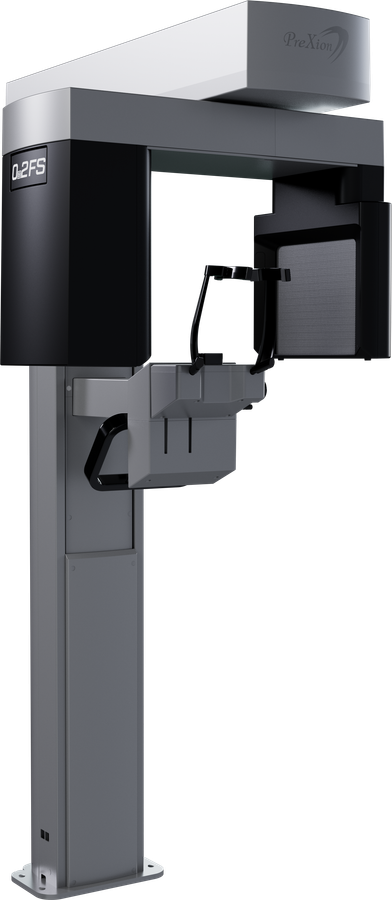
The Excelsior Endo is PreXion's dedicated high-resolution scanner, purpose-built for the detail endodontics demands: a 0.2mm focal spot — smaller than the 0.5mm spot on the rest of the Excelsior line — combined with voxels as small as 48 µm, the finest resolution PreXion offers in a dental CBCT. Where the Excelsior Mid and Max are built for full-arch coverage, with fields of view up to 16×9 cm and 16×21 cm and a 75 µm voxel floor, the Endo trades that larger field for resolution: its widest field of view tops out at 14×9 cm, and its smallest, 5×5 cm, is where the 48 µm mode lives — sized for a single tooth, implant site, or third molar rather than a full arch. A smaller focal spot means less geometric blurring, so images hold higher resolution than a conventional focal spot produces — the difference shows up in fine root fracture lines and accessory canal anatomy that a 0.5mm-spot scanner tends to smooth over. The Excelsior Endo also uses a horizontal beam source, which reduces the exam's dependence on metal-artifact-reduction algorithms in the first place rather than just correcting for the artifacts afterward — a useful property in a unit that spends much of its life imaging teeth that already carry posts, obturation material, or existing metal restorations. The Endo is a single-purpose tool, not a general-practice replacement for a panoramic or full-arch unit. It has no panoramic, bitewing, or cephalometric modes, and no 16 cm-class full-arch field of view. What it does, it does at a resolution nothing else in PreXion's dental line matches — the tradeoff an endodontic-focused practice is making when it chooses the Endo over the Mid or Max.
New to CBCT, or narrowing down field of view, voxel size, and cost? Read the complete guide to dental CBCT before you decide.
New: the Excelsior Endo deep dive
See how the 0.2 mm focal spot, 48 µm Endo mode, motion correction, metal control, flexible fields of view, and support model come together.
Under the hood
A smaller focal spot means less geometric blurring, so the beam produces a sharper, more detailed image than a conventional 0.5mm-spot scanner — the same principle that makes a smaller aperture sharpen a photograph. Paired with the Excelsior Endo's 48 µm voxel size, it's what lets the scanner resolve fine root fracture lines, accessory canals, and other endodontic detail that a general-purpose CBCT's larger focal spot tends to smooth over.
Patients make small, involuntary movements during a scan, and at endodontic resolution even a fraction of a millimeter of motion can blur a canal. PreXion's PMC algorithm detects and corrects for that movement automatically, so exams come out diagnostic-quality on the first pass instead of requiring a retake and a second exposure.
Root canal cases routinely involve gutta-percha, posts, or existing metal restorations, all of which throw off streaking artifacts that can obscure the anatomy being scanned. The Excelsior Endo's 3-level MAR corrects for that streaking, and because it's a reprocessing step rather than a rescan, a different correction level can be tried on the same scan without exposing the patient again.
Most of the burden of dealing with metal artifacts falls on MAR software after the fact. The Excelsior Endo's horizontal beam geometry reduces how much artifact the metal introduces in the first place, cutting the exam's reliance on MAR correction rather than just papering over the problem afterward.
The Excelsior Endo's base acquisition software is built around endodontic capture, not implant workflow — planning tools live in the optional OnDemand3D module instead. It adds 3D implant positioning and simulation, virtual endoscopy for exploring canal and airway anatomy from inside the reconstructed volume, STL conversion for digital surgical guide design and 3D printing, DICOM fusion to align and overlay multiple scans, and exportable 3D reports in HTML, PPT, or PDF.
Sample captures
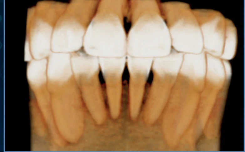
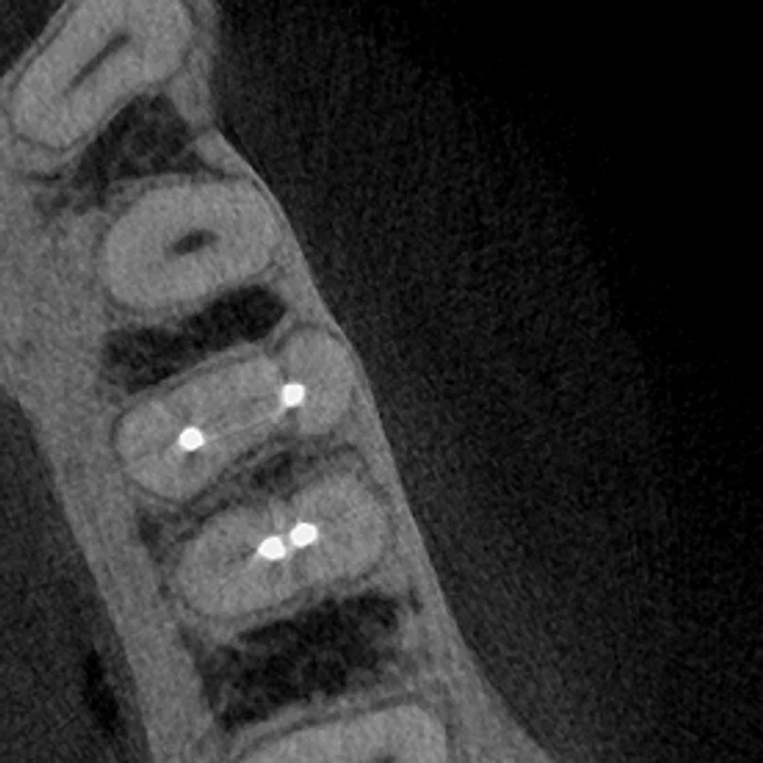
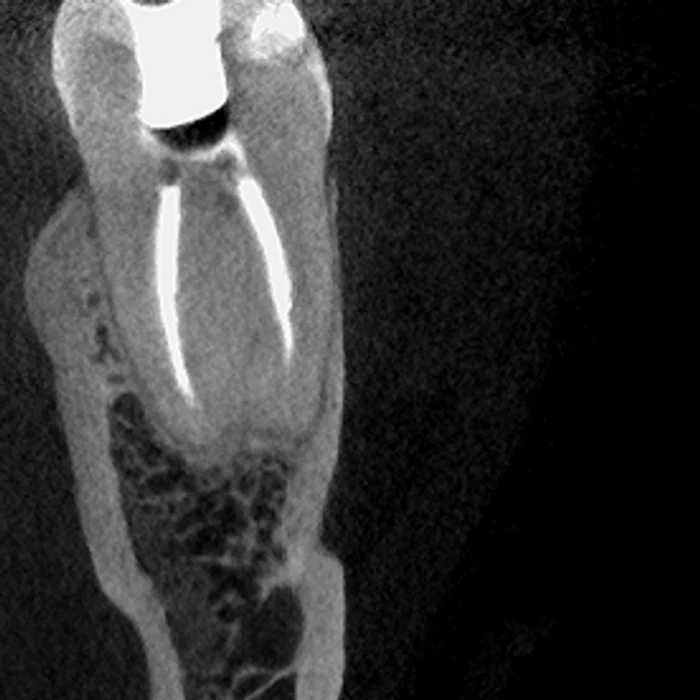
Actual radiographs produced by this system — the image quality you're buying, not a brochure rendering.
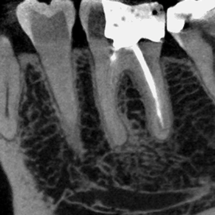
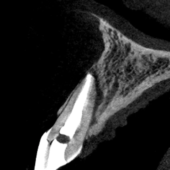
Its focal spot: 0.2mm versus the 0.5mm on the Excelsior Mid and Max. A smaller focal spot means less geometric blurring, so the Excelsior Endo resolves finer detail — down to a 48 µm voxel — which is why PreXion built it specifically for endodontic diagnosis.
Four fields of view: 5×5 cm, 9×6 cm, 9×9 cm, and 14×9 cm — the 14×9 cm option is described by PreXion as an extended full-arch field covering the third molar and zygomatic arch regions in a single volume.
Yes. It uses a horizontal beam source, which reduces the exam's dependence on metal-artifact-reduction algorithms in the first place, on top of the 3-level Metal Artifact Reduction (MAR) software also included.
No — ceph is not offered on the Excelsior Endo configuration; it's a dedicated high-resolution endodontic scanner rather than a general-purpose or ceph-capable unit.
The Excelsior Endo runs Software Eye with 3-level MAR. Every scan also exports in the standard DICOM format, so it's compatible with any DICOM-compliant viewing or planning software your practice already uses, not just Software Eye.
Every PreXion Excelsior Endo comes with manufacturer-backed installation and support from PreXion. Dental TI adds another layer on top: site planning, integration with your practice's imaging software, on-site team training with remote training also available, and expert remote and on-site support of our own. Dental TI has served dentistry since 2003 from Carmel, Indiana.
No. Even with the horizontal beam source already reducing reliance on artifact correction, the Excelsior Endo's 3-level Metal Artifact Reduction can reprocess the same scan at a different correction level after the fact — for gutta-percha, posts, or metal restorations — without a new exposure.
The scanner head uses an aeronautical-grade injected-aluminum housing with cooling fins and two high-flow, low-noise fans, keeping the system running at its rated resolution without thermal throttling during a busy schedule.
Because of its higher-resolution imaging, the Excelsior Endo calls for more than PreXion's standard 3D workstation spec: Windows 10 64-bit Professional, Intel Core i7 (10th gen or newer, 12 MB cache, 4.0 GHz+), 32 GB RAM, an Nvidia GeForce RTX 4070 12GB or better, a 500W+ power supply, 1TB+ storage, a 1920×1080 monitor, and a dedicated Gigabit Ethernet adapter.
Yes — PreXion requires equipment assembly to be performed by a certified technician, and the shipping packaging must stay sealed until they arrive; opening it early voids the manufacturer's warranty. Dental TI coordinates this as part of installation.
Endodontic diagnosis depends on resolving fine detail — accessory canals, small fracture lines, subtle periapical changes — at a scale where a conventional focal spot introduces enough geometric blurring to hide it. The Excelsior Endo's 0.2mm focal spot, less than half the 0.5mm spot on the rest of the Excelsior line, is paired with a voxel size as small as 48 µm specifically so that level of detail survives into the reconstructed image.
The Excelsior Endo goes finer: its voxel size runs from 48 to 200 µm, versus a 75 to 400 µm range on both the Mid and Max. That's because the Mid and Max are built for full-arch coverage — fields of view up to 16×9 cm on the Mid and 16×21 cm on the Max — while the Endo trades that larger field, topping out at 14×9 cm, for the sharpest image PreXion's dental line produces.
Yes, through the optional OnDemand3D software rather than the unit's base Eagle Eye acquisition software. OnDemand3D adds 3D implant positioning and simulation, along with virtual endoscopy, STL conversion, DICOM fusion, and exportable 3D reports — the same planning module available across PreXion's CBCT line.
It corrects for the small, involuntary movements patients make during a scan. At the Excelsior Endo's resolution, even minor motion can blur fine canal detail, so PMC's automatic correction is what keeps a high-resolution exam diagnostic-quality without needing a retake.
No. It's a dedicated CT scanner with no panoramic, bitewing, or cephalometric modes — everything about the Excelsior Endo is built around producing the highest-resolution 3D volume PreXion offers, not replacing a practice's 2D imaging unit.
Tell us your case mix, room, imaging software, and timing. We'll map the right configuration and build a complete estimate with the system, installation, integration, and training.