
Meet the PreXion Excelsior Endo: The CBCT Built Around Endo Detail
· David Hanning
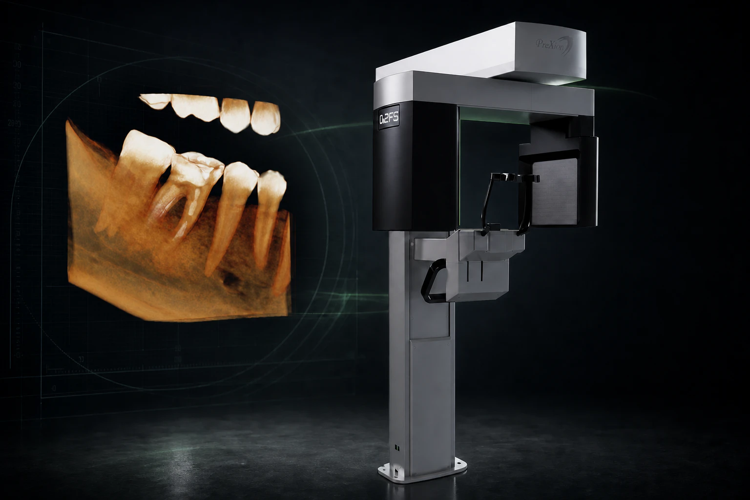
Most CBCT launches start with more: more field of view, more programs, more ways to make one machine serve every department.
The new PreXion Excelsior Endo starts with less.
Less blur at the X-ray source.
Its 0.2 mm focal spot is the smallest in the industry. Paired with a 48 µm voxel mode inside a focused 5 x 5 cm field, you get phenomenal preservation of fine anatomy.
It’s not just a badge—this machine is built from the ground up to optimize endodontic applications. In this post, we will review the many features that make this machine a top choice for endodontic practice.
See the Excelsior Endo, review actual scans, and request pricing →
The tightest focal spot in the category (0.2 mm)
CBCT buyers hear about voxel size constantly. Voxel size describes the reconstructed grid, but it doesn’t tell you how much geometric blur entered the image while the X-ray was being produced.
The focal spot is the small area on the tube target from which the X-rays originate. A larger source behaves like a broader light: edges spread. A smaller source narrows that geometric unsharpness before the detector and reconstruction algorithms get involved.
The Excelsior Endo’s 0.2 mm focal spot is 60% smaller in nominal dimension than the 0.5 mm spot used in the Excelsior Mid and Max. Then, in its focused 5 x 5 cm Endo mode, the Endo samples the reconstructed volume at an isotropic voxel size as small as 48 µm. The focal spot protects the detail entering the imaging chain; the voxel grid gives the reconstruction room to represent it.
A tiny voxel cannot recover detail that arrived blurred. That is why the two numbers belong together.
No specification guarantees that every crack or accessory canal will be visible—positioning, exposure selection, patient movement, metal, reconstruction, display, and clinical interpretation all still matter. But starting with a narrower X-ray source gives the rest of the imaging chain better information to work with.
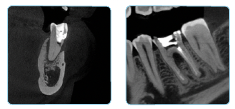
PreXion designed the rest of the system around the cases that fight back
Endodontic imaging rarely happens in perfect conditions. Patients move. Existing treatment and restorations introduce streaking. The anatomy you need may sit next to gutta-percha, a post, an implant, or dense metal.
The Excelsior Endo answers those problems at several points in the imaging chain:
-
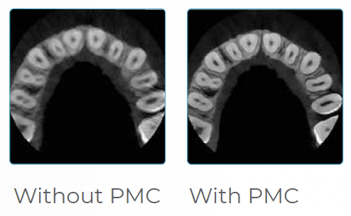
Patient Motion Correction (PMC): PreXion’s reconstruction algorithm is designed to correct image imperfections caused by patient micro-movement, protecting a high-resolution exam from the small motion that can soften canal detail.
-
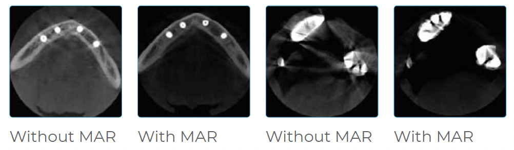
Metal Artifact Reduction from 0 through 3: The team can compare the original reconstruction with three selectable correction levels. If a different level is more useful, the same acquisition can be reprocessed without exposing the patient again.
-
A horizontal beam source: The dedicated horizontal geometry reduces the system’s dependence on metal-artifact-reduction algorithms in the first place. That matters in an Endo population where posts, obturation material, and restorations are normal, not edge cases.
-
LD, STD, HD, and UHD protocols: The operator can choose the resolution and exposure approach around the diagnostic task rather than scanning every patient at the maximum setting.
-
A thermally managed scanner head: The housing uses injected aluminum with cooling fins plus two high-flow, low-noise fans to move heat away from the acquisition system during the workday. PreXion has always been an extremely reliable brand for our clients, and it continues to innovate on technologies that keep its units scanning for a decade or more.
-
Three-axis scanner movement: Two orthogonal movements plus rotation give the system more flexibility in how it creates radiographic profiles, optimizes the slice plane, and maintains consistent vertical magnification.
-
A connected software workflow: The software package includes one server and five client licenses, with patient and exam management, TWAIN acquisition, DICOM import/save/send, PACS communication, DICOM worklist, viewer creation, measurements, annotations, and reporting. Optional OnDemand3D extends the system into implant planning, STL conversion, DICOM fusion, virtual endoscopy, and custom 3D reports.
The official Excelsior Endo brochure documents the focal spot, voxel range, FOVs, processing levels, cooling design, and system requirements in detail.
Four fields of view keep a focused machine from becoming a one-case machine
The 5 x 5 cm field is the headline because that is where the 48 µm mode lives. It is built around a focused diagnostic question: a tooth, an implant site, a third molar, and the anatomy immediately around it.
But this unit can also cover larger general and restorative workflows. Three other FOVs keep the system useful beyond a single-tooth volume:
| Field of view | What it covers |
|---|---|
| 5 x 5 cm | Focused Endo, local diagnosis, a single implant site, or a third molar |
| 9 x 6 cm | One arch or one TMJ region |
| 9 x 9 cm | Full arch, including maxilla, mandible, and ramus |
| 14 x 9 cm | Extended full arch through the third-molar and zygomatic regions |
One important limitation for our general practices: it has no panoramic, bitewing, or cephalometric modes. If your practice needs routine 2D imaging, a 16 cm field, or future ceph expansion, compare the Excelsior Mid or Max. If the priority is the most focused acquisition system in the line, the Endo is the platform to investigate.
It fits the direction Endo imaging is moving
The 2025 AAE/AAOMR position statement says CBCT should be used selectively, not routinely, with protocols that are indication- and patient-specific. It emphasizes the ALADAIP principle: exposure should be as low as diagnostically acceptable for the particular patient and task. The AAE’s summary of the updated guidance also puts training and interpretation alongside the hardware.
The Excelsior Endo gives the operator four field sizes and four resolution modes so the exam can be built around the question. Our team at Dental TI then trains your staff on field selection, positioning, capture, reconstruction, export, and the responsibility to interpret the entire acquired volume.
World-class reliability is hardware plus the people who own the handoff
PreXion has been specializing in dental 3D imaging since 2007 and maintains an awesome, dedicated U.S. support team.
Dental TI adds an additional layer that turns this great platform into a working system inside your office:
- site and room planning before the unit ships;
- certified equipment installation;
- workstation, network, TWAIN, DICOM, and imaging-software integration;
- on-site team training, with remote training also available; and
- expert remote and on-site support after go-live.
That is what reliability looks like in the real world: the scanner is engineered to run, the workflow is engineered to fit, and expert support is a phone call away when the day does not go according to the brochure.
The right next step is an estimate built around your cases
If you are seriously evaluating CBCT for Endo, do not stop at a brochure download.
Bring us the cases you refer out, the 2D and 3D equipment you already own, the imaging software your team uses, the room you have available, and the timing you are working toward. We will tell you whether the Excelsior Endo fits, where the Mid or Max makes more sense, and what the complete installed system should include.
Then we will put real numbers around it: equipment, software, installation, integration, training, and the options your workflow actually needs. Plus, we can show real patient volumes and get you confident in navigating them before the unit even arrives.
Review the Excelsior Endo and request your consultation and pricing →
Connect with us to learn exactly what it would take to put this sort of scan quality to work in your practice.