
What’s New With HDX WILL CBCT: Better Reconstruction and Smarter Workflow
· David Hanning
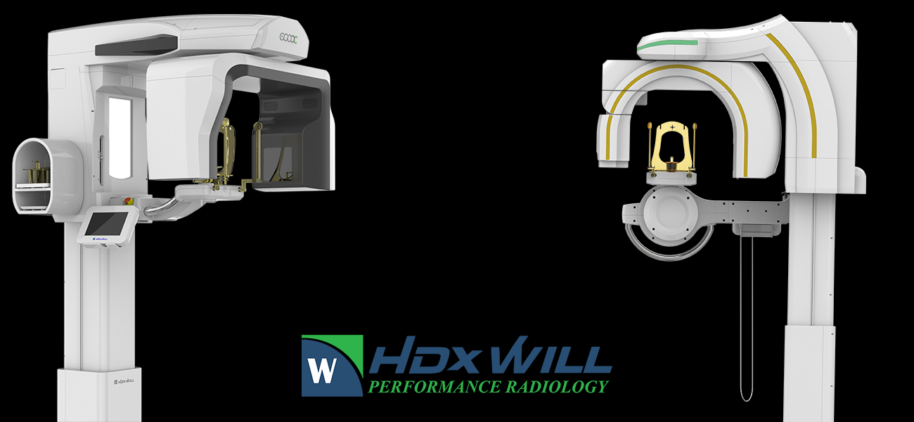
When most people compare CBCT systems, they start with the hardware: detector, voxel size, field of view, and footprint. Those things matter. But HDX WILL’s latest story is also about what happens after the scan.
Its current imaging platform combines an enhanced reconstruction engine, updated metal artifact reduction, flexible scan volumes, and software tools that can shorten the path from acquisition to planning. That makes this a useful time to look at what has changed and what those updates can mean in a working dental practice.
The reconstruction engine is the center of the update
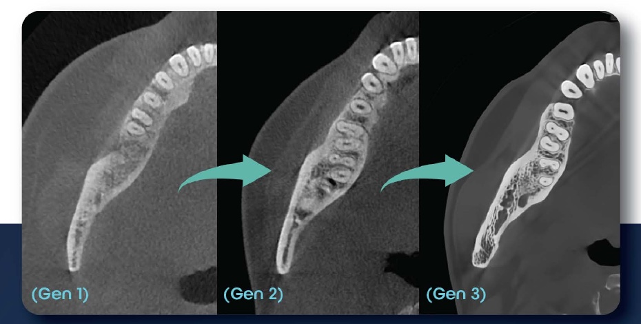
HDX WILL calls its ongoing image-reconstruction development CYOPTIX. The company describes it as a software effort that evaluates voxel data to improve contrast and sharpness, and its current brochure shows the progression across three generations of reconstruction.
That distinction matters. A CBCT image is not simply a picture taken by the detector. The system has to reconstruct the acquired data into a volume that a clinician can navigate. Changes in reconstruction can affect noise, edge definition, scatter, and the visibility of fine anatomy.
CYOPTIX keeps evolving, with reconstruction improvements that bring sharper detail and clearer images to diagnosis and treatment planning. For practices with compatible systems, those updates can help extend the useful clinical life of the hardware, so your HDX WILL investment keeps delivering value as the software advances.
See HDX WILL’s reconstruction overview
Next-generation metal artifact reduction
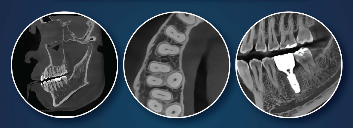
Metal restorations, implants, posts, and other dense materials can create streaking and shading that obscure nearby anatomy. HDX WILL’s current brochure describes a next-generation metal artifact reduction algorithm, while the company’s technology page identifies the feature as PrecisionMAR.
The goal is straightforward: reduce scatter and beam-hardening artifacts so the structures around metal are easier to inspect. That is useful in implant planning, endodontic evaluation, oral surgery, and any case where existing dentistry sits close to the anatomy in question.
Artifact reduction should still be judged on the cases you expect to scan. No algorithm removes every acquisition problem, and the right field of view, exposure, positioning, and patient stability still matter.
One platform, from a focused 3 × 3 cm volume to broader anatomy
The Eco-X and Dentri Max share the same basic imaging and software approach, but cover different upper ends of the field-of-view range.
- Eco-X: free field-of-view selection begins at 3 × 3 cm and extends through 12 × 9 cm, with fixed modes up to 16 × 9 cm. Its published voxel options include 70, 100, 150, and 200 µm.
- Dentri Max: begins with the same focused 3 × 3 cm selection and adds broader modes, including 16 × 9 cm and a maximum 18 × 16.5 cm field for larger maxillofacial studies.
That range lets a practice select the anatomy required for the case instead of using one large volume for everything. A focused endodontic study, full-arch implant plan, airway review, and maxillofacial case do not need the same capture.
Review the Eco-X specifications or the Dentri Max specifications.
Workflow tools go beyond the 3D viewer
The current HDX WILL platform includes tools for several jobs that often require extra steps:
- FMX extraction from a panoramic image can build a familiar series of views from one panoramic acquisition.
- Model scan mode can produce CBCT DICOM data and an STL file from a stone model, impression, or denture.
- OnDemand3D supports 3D navigation, implant planning, airway analysis, and 3D cephalometric work.
- Will-Master manages patient images and 2D viewing.
- WillCeph adds automatic landmark tracing, analysis, treatment simulation, reporting, and image superimposition for practices using a cephalometric configuration.
The value is not that every practice will use every feature. It is that the system can support a broader workflow as the practice adds procedures or brings more planning in-house.
Explore HDX WILL’s specialty software
Dose control still starts with the patient and the protocol
HDX WILL’s Adaptive Exposure Control adjusts exposure based on patient anatomy and tissue thickness. The platform also offers low-dose modes and pediatric field options.
Those tools support a sound imaging principle: use the smallest volume and the lowest exposure that still answer the clinical question. The operator still has to choose the right protocol, position the patient well, and confirm that the scan will cover the anatomy needed for diagnosis.
Read HDX WILL’s dose-control overview
See the image data, not just the brochure
Specifications describe what a system can acquire. A real DICOM volume shows how the data feels to navigate.
Dental TI’s browser-based viewer includes anonymized Eco-X endodontic volumes that you can inspect in axial, coronal, and sagittal views. It is a useful first look before a live workstation demonstration, and it gives your team something concrete to discuss.
Open the HDX WILL Eco-X sample volumes
A practical education library for the team
HDX WILL’s clinical education does a good job of connecting imaging features to real diagnostic work. The available sessions cover image interpretation, endodontics, implants, airway, TMJ, orthodontics, and software technique. That makes the library useful after installation as well as during the buying process.
We have organized a short set of those videos by clinical topic below. Start with the work your practice already does, then use the adjacent topics to see where the system can support growth.
The seven-part series is hosted by Tim Todack of HDX WILL North America and moves from diagnosis into planning and follow-up:
Focused diagnosis
- 3D Endodontic Diagnosis, including OnDemand3D navigation and focused field-of-view selection
- 3D Bilateral TMJ Evaluation, including bilateral slice review
- 3D Airway Evaluation, including endoscopic and volumetric airway views
Planning and treatment
- Digital Implant Planning, including implant selection, measurement, and nerve mapping
- 3D Orthodontic Evaluation, including derived panoramic and cephalometric views
Digital workflow and follow-up
- Incorporating an STL File Into CBCT Diagnosis, showing an intraoral scan merged with CBCT data
- 3D Superimposition With CBCT, using before-and-after volume fusion to visualize treatment changes
Browse the official HDX WILL North America and HDX Academy channel
What Dental TI adds
The equipment is only one part of a successful CBCT project. Dental TI helps plan the room, coordinate installation, connect the imaging workflow, train the team, and provide expert support after go-live. Most software and workflow questions can be handled remotely, often without waiting for a service visit.
The best next step is to bring us three things: the procedures you expect to scan most, the largest anatomy you routinely need, and the software your team uses today. From there, we can show you the HDX WILL configuration and sample volumes that fit the conversation.