
A Dental X-Ray Retake Can Begin Before the Exposure
· David Hanning
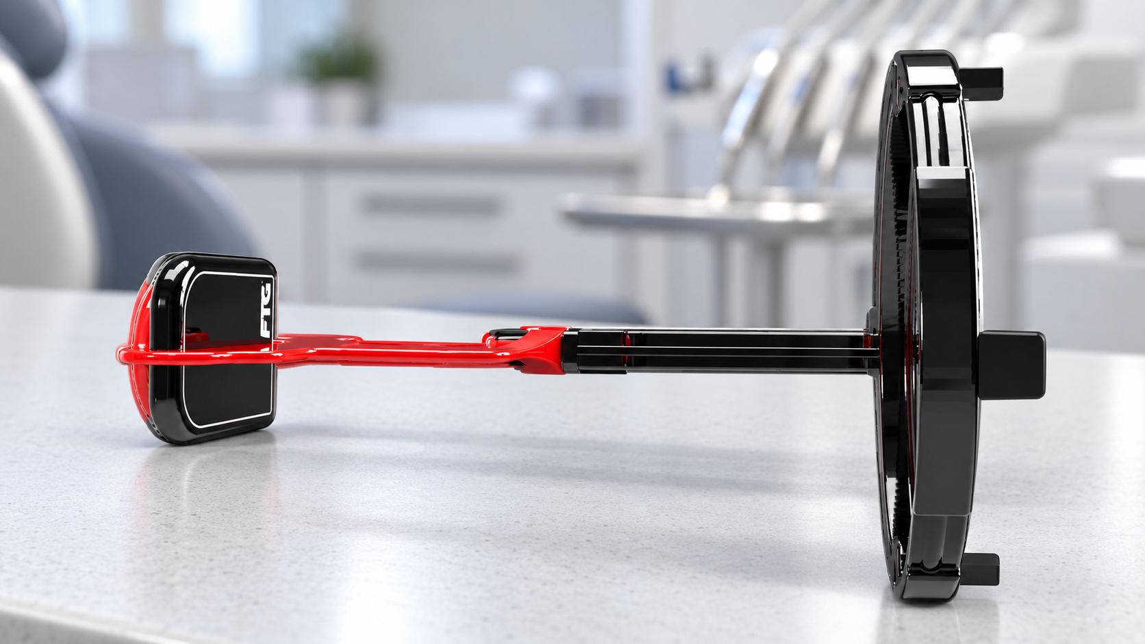
A retake does not always begin with the exposure setting.
Sometimes it begins while the assistant is still positioning the sensor.
The holder is aligned. The patient starts to close. The cable catches on the cheek, pulls against the ring, or limits the angle the assistant wanted. The final position is close enough to try, but not the position the team intended.
The exposure happens later. The cause arrived earlier.
Wireless capture does not eliminate every positioning error. It removes one specific force from the workflow: the cable.
The cable is part of the geometry
It is easy to think of the cable as a connection rather than a positioning variable. In the mouth, it is both.
The cable has weight and stiffness. It has to exit the mouth, travel around the patient, and reach a computer or interface. It can contact the holder, cheek, bib, chair, assistant, or patient. A tight bend near the strain relief can create its own resistance.
The effect may be small. In intraoral positioning, small changes can decide whether a contact opens, an apex is included, or the receptor stays parallel to the anatomy.
This does not mean every closed contact or cone cut came from the cable. Holder choice, patient anatomy, training, beam alignment, sensor size, and movement still matter. The point is that the cable belongs on the list of variables.
The retake cost is more than another image
When the first capture misses the intended anatomy, the team has to decide quickly:
- reposition and expose again;
- accept a limited image;
- change holders;
- move the patient or tube head;
- troubleshoot a capture that did not return; or
- borrow another room or sensor.
A retake adds time and another exposure. It can also change the patient’s confidence. The first exposure beep happened. Now the team has to explain why another image is needed.
At low frequency, each event feels trivial. Across thousands of images, the minutes, interruptions, and patient explanations become a real operating cost.
What changes when the sensor is not tethered
DC-Air has no sensor cable or USB tether during capture. The charging dock connects separately.
That changes positioning in several practical ways:
- nothing pulls from the sensor toward the computer;
- the assistant can approach the holder without routing a cable;
- the sensor can be oriented around the holder and anatomy without planning an exit path for the tether; and
- the cable-related failure mode is absent from the sensor itself.
The honest question is whether removing the cable improves positioning and repeatability for your team.
Test the workflow, not one perfect placement
A trade-show demonstration is not enough.
Build a short trial that includes the people and images the practice actually produces:
- Use the same operatories and X-ray sources.
- Include the assistants and hygienists who take most of the images.
- Compare common bitewings, periapicals, and challenging positions.
- Record placement time and repositioning before exposure.
- Record retakes and the reason for each one.
- Confirm that every capture reaches the imaging software reliably.
- Ask patients about comfort without turning one comment into a universal claim.
- Review native image detail separately from the positioning workflow.
The trial should answer two questions: does the sensor help the team place and capture the image, and does the resulting image contain the detail the clinician wants?
Do not trade one workflow problem for another
Wireless matters only if the rest of the system works.
Confirm:
- charge capacity and dock workflow;
- the number of sensors and rooms the practice needs;
- software integration;
- how the image is associated with the correct room and patient;
- training and support;
- warranty and protection options; and
- what happens if the sensor is dropped, damaged, or misplaced.
DC-Air is documented for 150 or more radiographs per charge and dock-based charging. Do not rely on an unapproved rapid-recharge claim. Build the charging routine around the practice’s actual image volume.
Positioning is one part of the complete comparison
A wireless sensor can remove cable drag while still producing an image the clinician does not prefer. A sharp-looking image can still rely on processing more than native detector detail. A strong detector can still disappoint if the software integration is weak.
Evaluate the complete chain:
- placement;
- exposure;
- detector capture;
- processing;
- display;
- software write-in;
- charging and room coverage; and
- support after go-live.
That is why Dental TI prefers a live comparison over a generic product pitch.
Bring one current sensor, the imaging software your team already uses, and the rooms you want to cover. We will put DC-Air beside the existing workflow, inspect the native images, and quote the setup the practice would actually need.