
The X-Ray Dial Doesn't Tell You the Dose. Here's Why
· David Hanning
Diagnostic Truth Series · Part 6
I’ve been in dental for 34 years, and I sold and supported digital X-ray systems for 26 of those years. For years, I sold a low-dose sensor. I rang the bell about the low exposure numbers, because that’s what I was handed to sell. I believed it.
Here’s what I didn’t say as loudly back then. The images were diagnostic. But they weren’t as sharp as the high-res sensors of the day. And those high-res sensors asked for meaningfully more exposure on the dial. The market didn’t care. Doctors who wanted the detail paid for it in a little more time on the timer and didn’t lose sleep over it. Fourteen years later, that high-res sensor is still on the wall. The low-dose sensor I sold? That company didn’t survive. The market ran the experiment with real dollars and picked the detail.
Here’s why.
The Number on the Dial Is Just a Timer
The number you set is on the X-ray head, and all it controls is exposure time. It only means something next to the other three settings: kV, mA, and how far the cone sits from the tooth. By itself, that timer number tells you almost nothing about the dose.
And distance matters more than people think. Move the cone an inch off the cheek, and the radiation reaching the sensor drops by more than the entire difference between a low timer setting and a high one. That’s the inverse square law. Same number on the dial, very different exposure.
There’s a patient-safety angle too. The end of the cone should sit right against the skin. Leave an air gap, and the beam keeps spreading across it, so the field landing on the patient is larger than necessary and captures tissue that doesn’t need imaging. Better still, run rectangular collimation. Sizing the field to just larger than the sensor cuts patient dose by about two-thirds. That’s the biggest dose lever you’ve got, and it costs nothing.
The Bench Data I Sat On for a Decade
Back in 2014, I consulted for a low-dose sensor manufacturer, comparing their production sensor to a higher-res one they were building. It was confidential then. That company is gone now, so here they are — the actual bench images, in microgray (µGy), which is the actual detector dose.
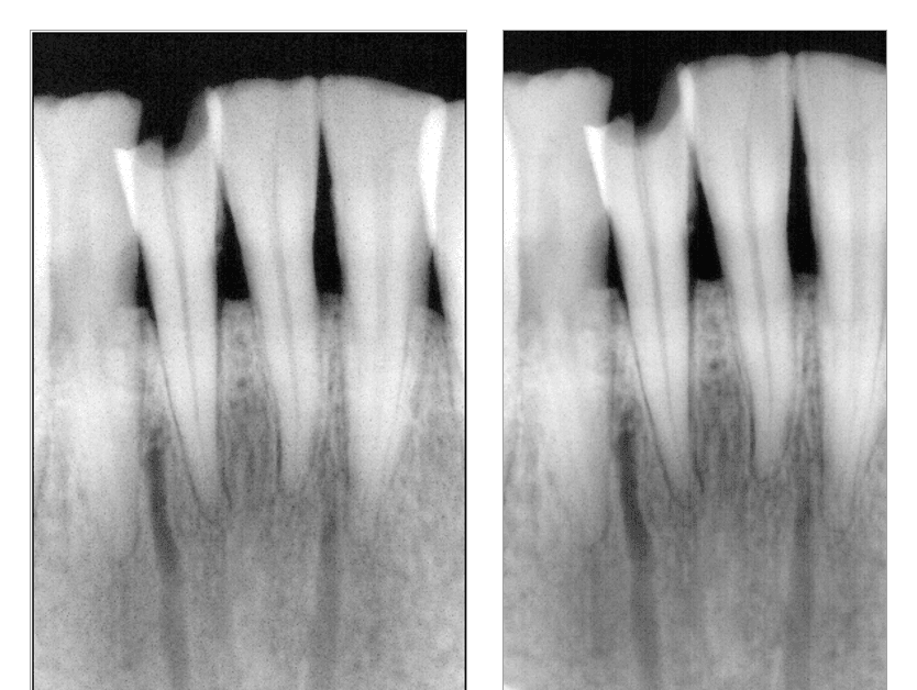 *The low-dose sensor at 80 µGy and 160 µGy — already at its ceiling. (Manufacturer bench images, 2014.)*
The sensor they sold as low-dose. At 80 µGy, and then at double the dose, 160: twice the dose, almost no new detail. The sensor was already at its ceiling. A low-res detector can’t turn extra photons into detail it isn’t built to resolve.
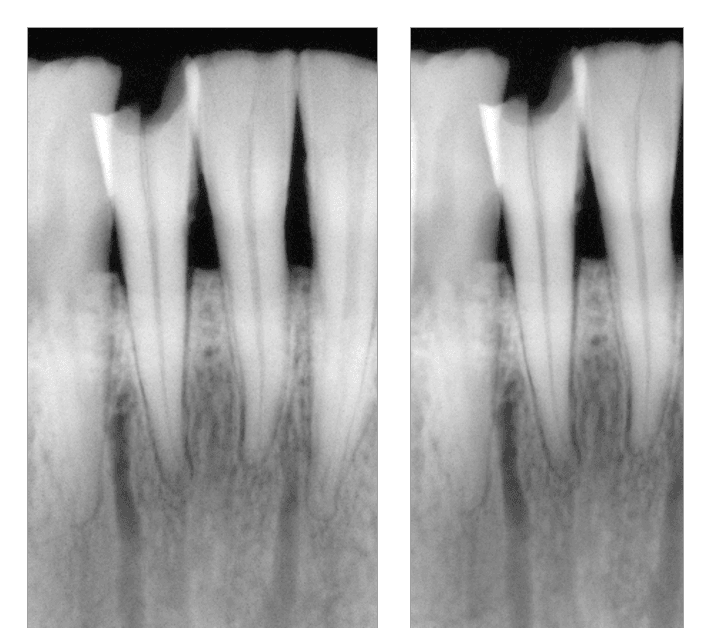 *The successor sensor at 160 µGy and 220 µGy — built to run at 2 to 3 times the original's 80 µGy baseline.*
That successor was the sensor built to replace the original low-dose one. In testing, we ran it at 160, then 220, near saturation. Read that again. The “low dose” company built its replacement to run at 2 to 3 times the dose of the sensor it replaced.
Because more resolution costs more exposure. There’s no way around it.
What Exposure Buys When the Detector Can Actually Resolve It
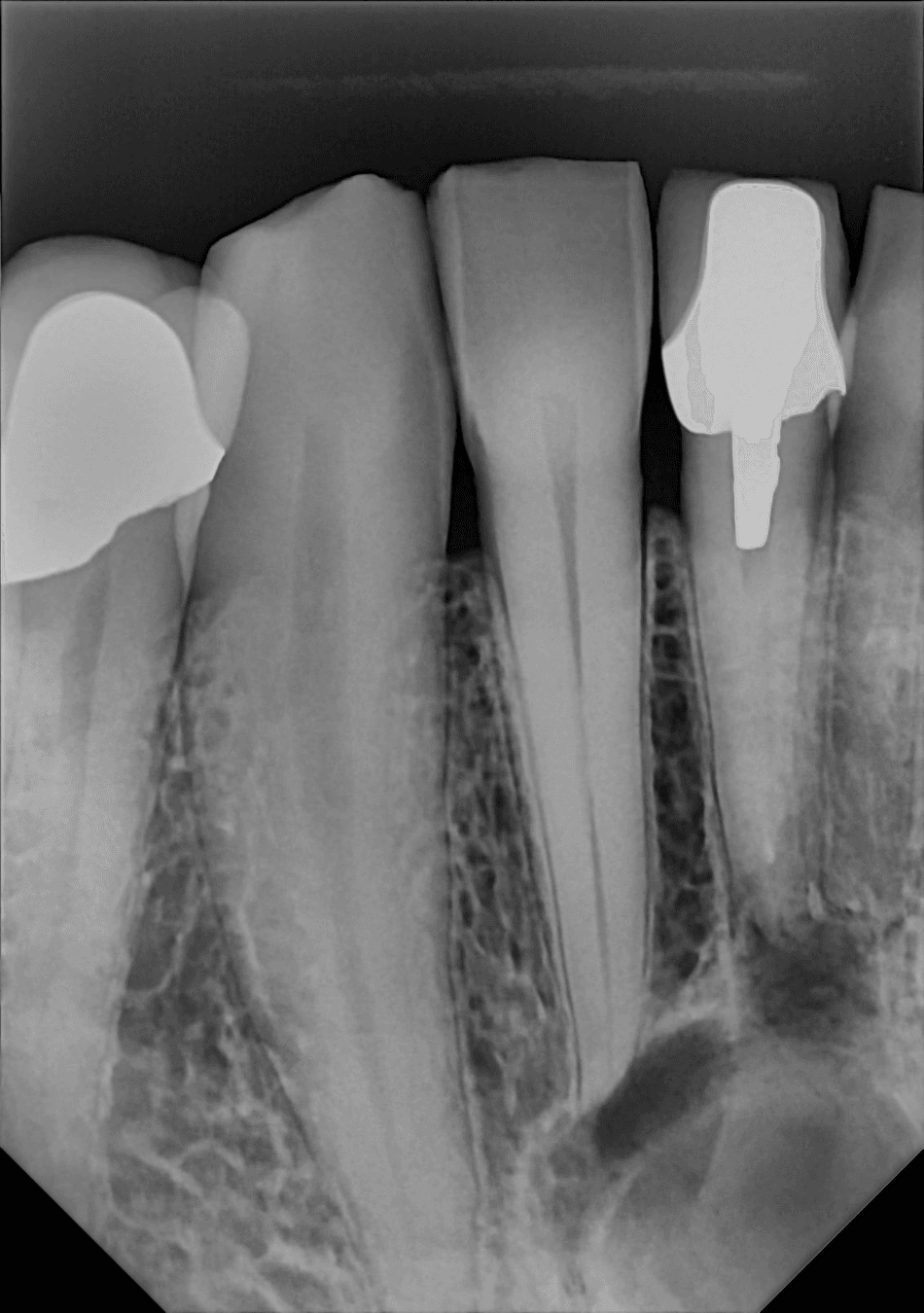
Now a modern high-res, direct-conversion sensor. Different patient, so read it for detail, not a head-to-head.
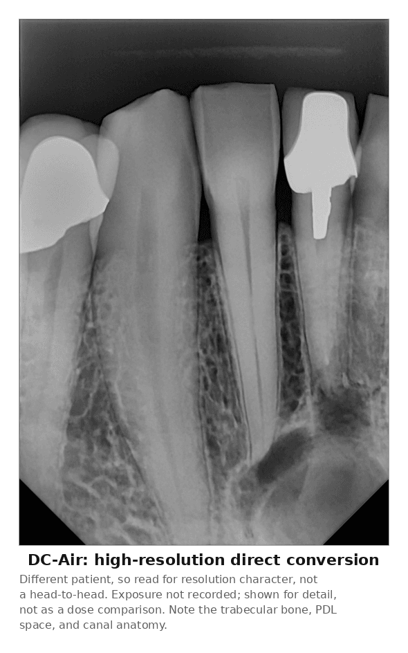 *A DC-Air™ direct-conversion capture — shown for resolution character, not as a dose comparison.*
Trabecular bone, PDL space, and canal anatomy. That’s what exposure buys when the detector can resolve it. Straight up: that’s a DC-Air™, and my company, Dental TI, distributes it. I don’t have the exposure logged on that shot, so take it as a resolution example, not a dose claim.
The numbers, if you want them. On a Gendex 770, that low-dose sensor ran at 3, 5, 7 pulses. A modern high-res sensor on that same 770 wants about 16, 20, 25. Sounds like a lot. It isn’t.
Put the Dose in Perspective
A digital intraoral is about a microsievert. Your patient gets roughly 8 µSv of background radiation every day just being alive. A cross-country flight is 30-plus. The gap between a low and a high timer setting is a rounding error to the patient.
And the standard isn’t “lowest dose” anymore. It moved to ALADA: as low as diagnostically acceptable. The newest version, ALADA-IP, adds two words that matter: indication-oriented and patient-specific. Match the dose to what you’re looking for and who’s in the chair. A dose so low you miss the diagnosis doesn’t pass that standard. It fails it.
You already do this on the CBCT. Low dose, low res for an airway. Turn it up for endo, because you need to resolve the canal. Nobody calls the endo scan reckless. The task sets the dose.
You Cannot Cheat Physics on a Native Image
Software can make a noisy, low-detail image look clean. It can’t put back detail that was never captured.
Want to actually lower dose? Technique. Ring to the skin, cone to the ring. Rectangular collimation. Good holders so you get it in one and never retake. That’s real dose reduction, and it costs you nothing.
The good news: you’ve got more room than the dose talk suggests. Every modern sensor, run the way it’s designed, sits well inside safe. So set your exposure for the clearest, most diagnostic image you can get, and feel good about it. Chase the detail. Your patients are better served by a film you can actually read.
I sold the low number for years, so I’ve seen this from both sides. I’d genuinely like to hear how you all weigh it. You read more films in a week than I do, so tell me where you land.
Technique note: the leading high-res sensor of that era carried a manufacturer guide calling for roughly 6 to 7 pulses (anterior) and 7 to 10 pulses (posterior) on a Gendex 770, versus the low-dose sensor’s 3, 5, and 7 — more exposure for more resolution, straight from the manufacturer.
Want to see a direct-conversion capture on your own toughest case? Book a walkthrough with Dental TI — ask about current pricing and our summer demo program. Prefer to watch it first? See the DC-Air™ video library.